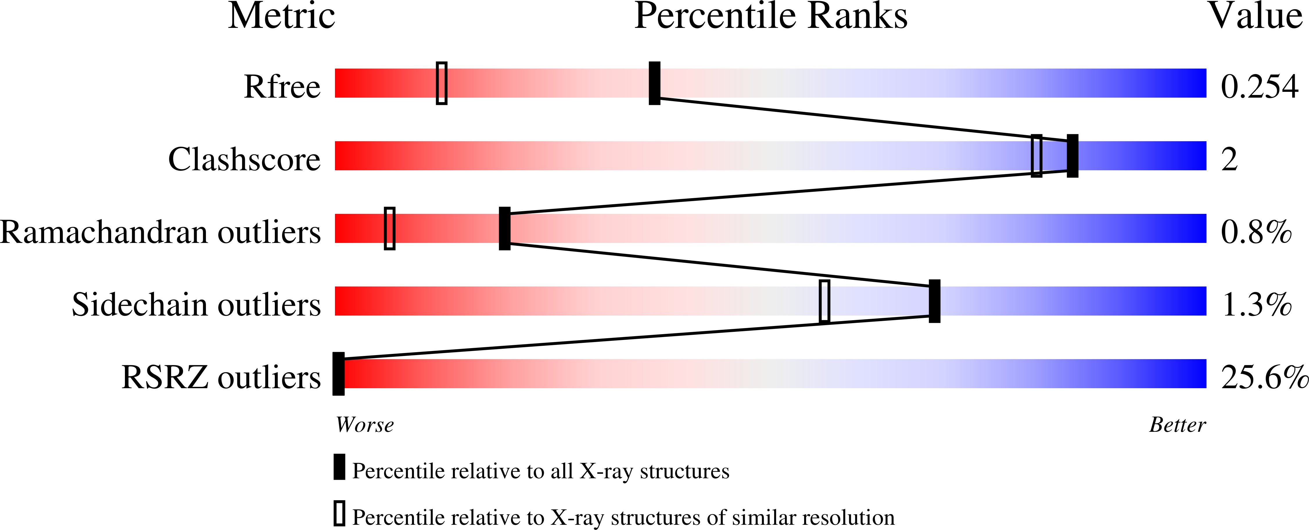
2VOA image
Deposition Date 2008-02-11
Release Date 2008-12-09
Last Version Date 2023-12-13
Entry Detail
PDB ID:
2VOA
Keywords:
Title:
Structure of an AP Endonuclease from Archaeoglobus fulgidus
Biological Source:
Source Organism:
Host Organism:
Method Details:
Experimental Method:
Resolution:
1.70 Å
R-Value Free:
0.23
R-Value Work:
0.19
R-Value Observed:
0.19
Space Group:
P 1 21 1
Macromolecular Entities
Polymer Type:polypeptide(L)
Molecule:EXODEOXYRIBONUCLEASE III
Gene (Uniprot):AF_0580
Mutations:YES
Chain IDs:A, B
Chain Length:257
Number of Molecules:2
Biological Source:ARCHAEOGLOBUS FULGIDUS
Polymer Type:polydeoxyribonucleotide
Molecule:5'-D(*GP*CP*GP*GP*TP*AP*GP*CP*CP*GP)-3'
Chain IDs:C
Chain Length:10
Number of Molecules:1
Biological Source:synthetic construct
Polymer Type:polydeoxyribonucleotide
Molecule:5'-D(*CP*GP*GP*CP*TP*AP*CP*CP*GP*CP)-3'
Chain IDs:D
Chain Length:10
Number of Molecules:1
Biological Source:synthetic construct
Modified Residue
Compound ID Chain ID Parent Comp ID Details 2D Image
OCS A CYS CYSTEINESULFONIC ACID
Primary Citation
Structure and Function of the Abasic Site Specificity Pocket of an Ap Endonuclease from Archaeoglobus Fulgidus.
DNA Repair 8 219 ? (2009)
PMID: 19015049 DOI: 10.1016/J.DNAREP.2008.10.009

Abstact

The major AP endonuclease in Escherichia coli Exonuclease III (ExoIII) is frequently used in gene technology due to its strong exonucleolytic activity. A thermostabilized variant of ExoIII or a homologous enzyme from thermophilic organisms could be most useful for further applications. For this purpose we characterized a nuclease from the hyperthermophilic archaeon Archaeoglobus fulgidus (Af_Exo), which shares 33% overall sequence identity and 55% similarity to ExoIII. The gene coding for this thermostable enzyme was cloned and expressed in E. coli. The purified protein shows a strong Mg(2+)-dependent nicking activity at AP-sites, nicking of undamaged double-stranded (ds) DNA and a weak exonucleolytic activity. A V217G variant of the enzyme was crystallized with decamer ds-DNA molecule, and the three-dimensional structure was determined to 1.7A resolution. Besides our goal to find or produce a thermostable exonuclease, the structural and catalytic data of Af_Exo and a series of mutant proteins, based on the crystal structure, provide new insight into the mechanism of abasic site recognition and repair. Each of the hydrophobic residues Phe 200, Trp 215 and Val 217, forming a binding pocket for the abasic deoxyribose in Af_Exo, were mutated to glycine or serine. By expanding the size of the binding pocket the unspecific endonucleolytic activity is increased. Thus, size and flexibility of the mostly hydrophobic binding pocket have a significant influence on AP-site specificity. We suggest that its tight fitting to the flipped-out deoxyribose allows for a preferred competent binding of abasic sites. In a larger or more flexible pocket however, intact nucleotides more easily bind in a catalytically competent conformation, resulting in loss of specificity. Moreover, with mutations of Phe 200 and Trp 215 we induced a strong exonucleolytic activity on undamaged DNA.

Legend

Protein

Chemical

Disease

Primary Citation of related structures