2VNV image
Deposition Date 2008-02-07
Release Date 2008-04-08
Last Version Date 2024-05-08
Entry Detail
PDB ID:
2VNV
Title:
Crystal structure of BclA lectin from burkholderia cenocepacia in complex with alpha-methyl-mannoside at 1.7 Angstrom resolution
Biological Source:
Source Organism:
Host Organism:
Method Details:
Experimental Method:
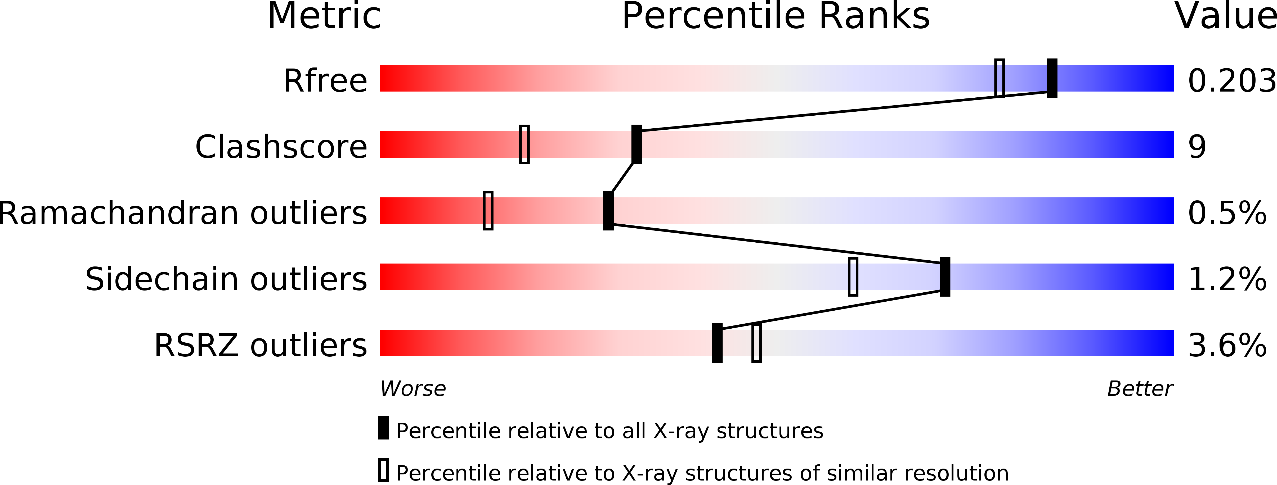
Resolution:
1.70 Å
R-Value Free:
0.20
R-Value Work:
0.16
R-Value Observed:
0.17
Space Group:
C 2 2 21
Macromolecular Entities
Polymer Type:polypeptide(L)
Molecule:BCLA
Chain IDs:A, B, C, D, E
Chain Length:129
Number of Molecules:5
Biological Source:BURKHOLDERIA CENOCEPACIA
Primary Citation
Structural Basis for Mannose Recognition by a Lectin from Opportunistic Bacteria Burkholderia Cenocepacia
Biochem.J. 411 307 ? (2008)
PMID: 18215132 DOI: 10.1042/BJ20071276

Abstact

Chronic colonization of the lungs by opportunist bacteria such as Pseudomonas aeruginosa and members of the Bcc (Burkholderia cepacia complex) is the major cause of morbidity and mortality among CF (cystic fibrosis) patients. PA-IIL (lecB gene), a soluble lectin from Ps. aeruginosa, has been the subject of much interest because of its very strong affinity for fucose. Orthologues have been identified in the opportunist bacteria Ralstonia solanacearum, Chromobacterium violaceum and Burkholderia of Bcc. The genome of the J2315 strain of B. cenocepacia, responsible for epidemia in CF centres, contains three genes that code for proteins with PA-IIL domains. The shortest gene was cloned in Escherichia coli and pure recombinant protein, BclA (B. cenocepacia lectin A), was obtained. The presence of native BclA in B. cenocepacia extracts was checked using a proteomic approach. The specificity of recombinant BclA was characterized using surface plasmon resonance showing a preference for mannosides and supported with glycan array experiments demonstrating a strict specificity for oligomannose-type N-glycan structures. The interaction thermodynamics of BclA with methyl alpha-D-mannoside demonstrates a dissociation constant (K(d)) of 2.75 x 10(-6) M. The X-ray crystal structure of the complex with methyl alpha-D-mannoside was determined at 1.7 A (1 A=0.1 nm) resolution. The lectin forms homodimers with one binding site per monomer, acting co-operatively with the second dimer site. Each monomer contains two Ca2+ ions and one sugar ligand. Despite strong sequence similarity, the differences between BclA and PA-IIL in their specificity, binding site and oligomerization mode indicate that the proteins should have different roles in the bacteria.

Legend

Protein

Chemical

Disease

Primary Citation of related structures