2VN2 image
Deposition Date 2008-01-30
Release Date 2008-08-12
Last Version Date 2024-05-08
Entry Detail
PDB ID:
2VN2
Keywords:
Title:
Crystal structure of the N-terminal domain of DnaD protein from Geobacillus kaustophilus HTA426
Biological Source:
Source Organism:
Host Organism:
Method Details:
Experimental Method:
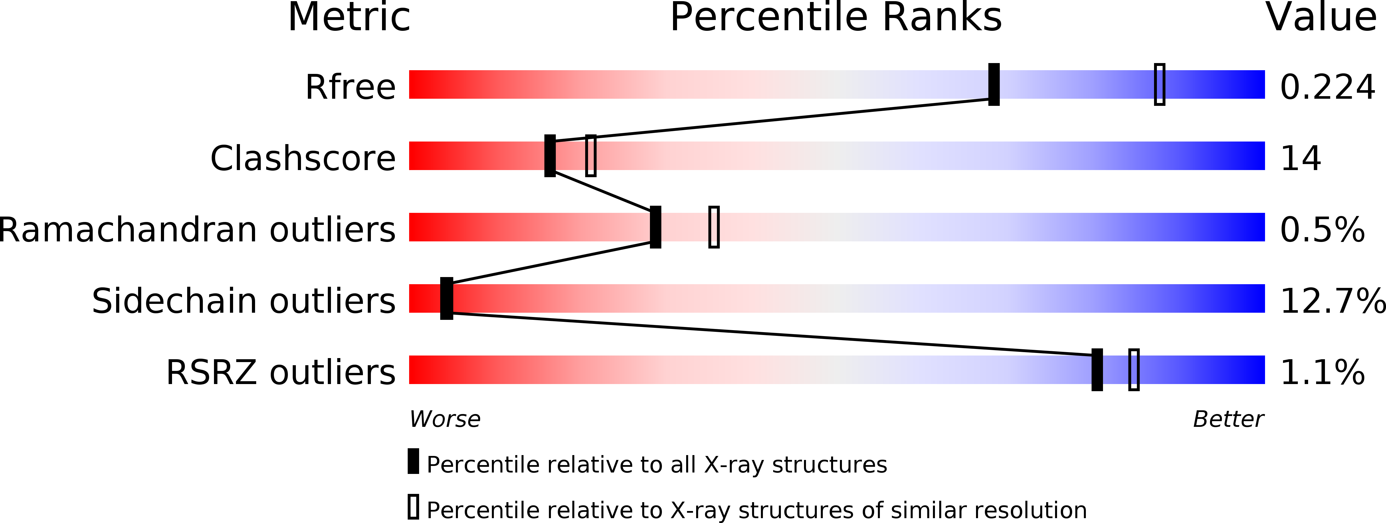
Resolution:
2.30 Å
R-Value Free:
0.22
R-Value Work:
0.21
R-Value Observed:
0.21
Space Group:
F 2 2 2
Macromolecular Entities
Polymer Type:polypeptide(L)
Molecule:CHROMOSOME REPLICATION INITIATION PROTEIN
Gene (Uniprot):dnaD
Chain IDs:A, B, C, D
Chain Length:128
Number of Molecules:4
Biological Source:GEOBACILLUS KAUSTOPHILUS HTA426
Ligand Molecules
Primary Citation
Crystal Structure of the N-Terminal Domain of Geobacillus Kaustophilus Hta426 Dnad Protein.
Biochem.Biophys.Res.Commun. 375 220 ? (2008)
PMID: 18703019 DOI: 10.1016/J.BBRC.2008.07.160

Abstact

The DnaD is one of the primosomal proteins that are required for initiation and re-initiation of chromosomal DNA replication in Gram-positive bacteria. The DnaD protein is composed of two major structural domains: an N-terminal oligomerization domain and a C-terminal ssDNA binding domain. Here, we report the crystal structure of the N-terminal domain (aa 1-128) of DnaD (DnaDn) of Geobacillus kaustophilus HTA426 at 2.3A resolution. The structure of DnaDn reveals an extended winged-helix fold, a typical double-stranded DNA binding motif as winged-helix proteins. DnaDn formed tetramers in the crystalline state, but the results of gel filtration chromatography further indicated that this domain of DnaD was a stable dimer in solution. The structural analysis of DnaDn may suggest the binding sites for DNA and DnaB, and an assembly mechanism for Gram-positive bacterial DNA replication primosome.

Legend

Protein

Chemical

Disease

Primary Citation of related structures