Deposition Date
2008-01-04
Release Date
2009-02-10
Last Version Date
2023-12-13
Entry Detail
PDB ID:
2VKY
Keywords:
Title:
Headbinding Domain of Phage P22 Tailspike C-Terminally Fused to Isoleucine Zipper pIIGCN4 (Chimera I)
Biological Source:
Source Organism(s):
ENTEROBACTERIA PHAGE P22 (Taxon ID: 10754)
SACCHAROMYCES CEREVISIAE (Taxon ID: 4932)
SACCHAROMYCES CEREVISIAE (Taxon ID: 4932)
Expression System(s):
Method Details:
Experimental Method:
Resolution:
2.05 Å
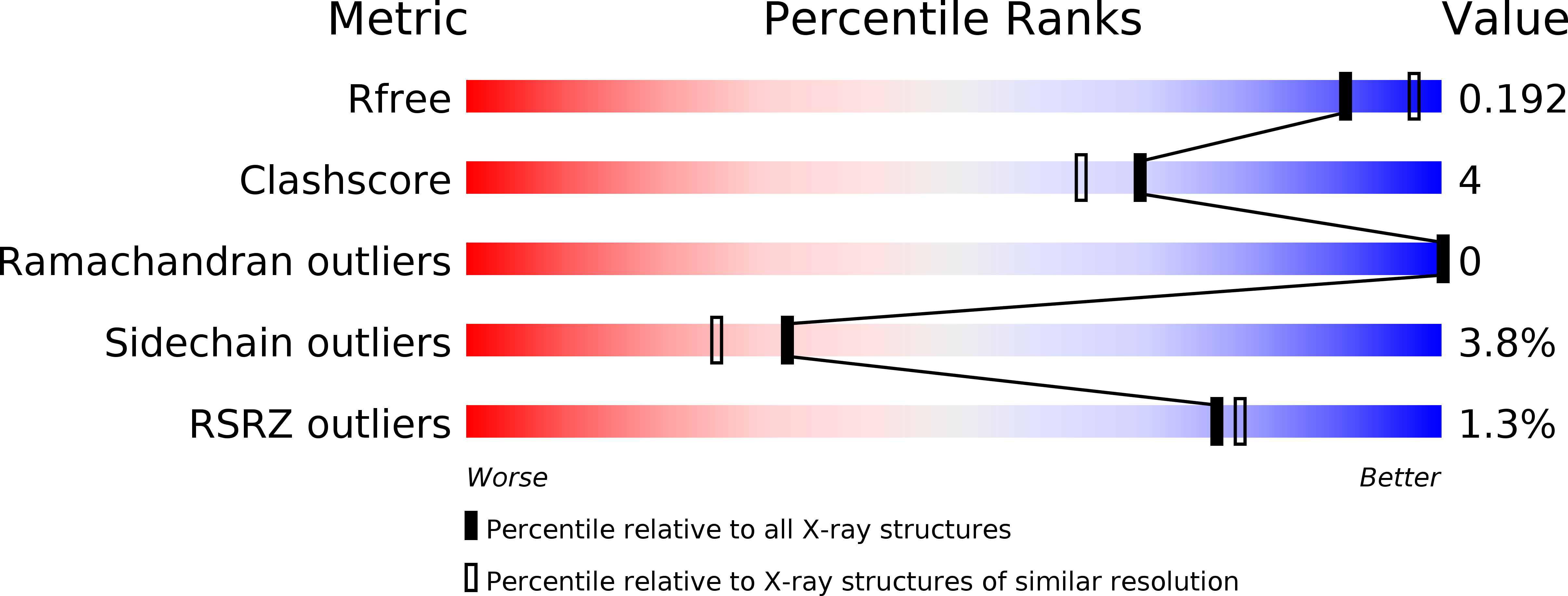
R-Value Free:
0.18
R-Value Work:
0.14
R-Value Observed:
0.14
Space Group:
P 63