Deposition Date
2007-12-18
Release Date
2008-03-18
Last Version Date
2024-05-08
Entry Detail
PDB ID:
2VKD
Keywords:
Title:
CRYSTAL STRUCTURE OF THE CATALYTIC DOMAIN OF LETHAL TOXIN FROM CLOSTRIDIUM SORDELLII IN COMPLEX WITH UDP-GLC AND MANGANESE ION
Biological Source:
Source Organism(s):
CLOSTRIDIUM SORDELLII (Taxon ID: 1505)
Expression System(s):
Method Details:
Experimental Method:
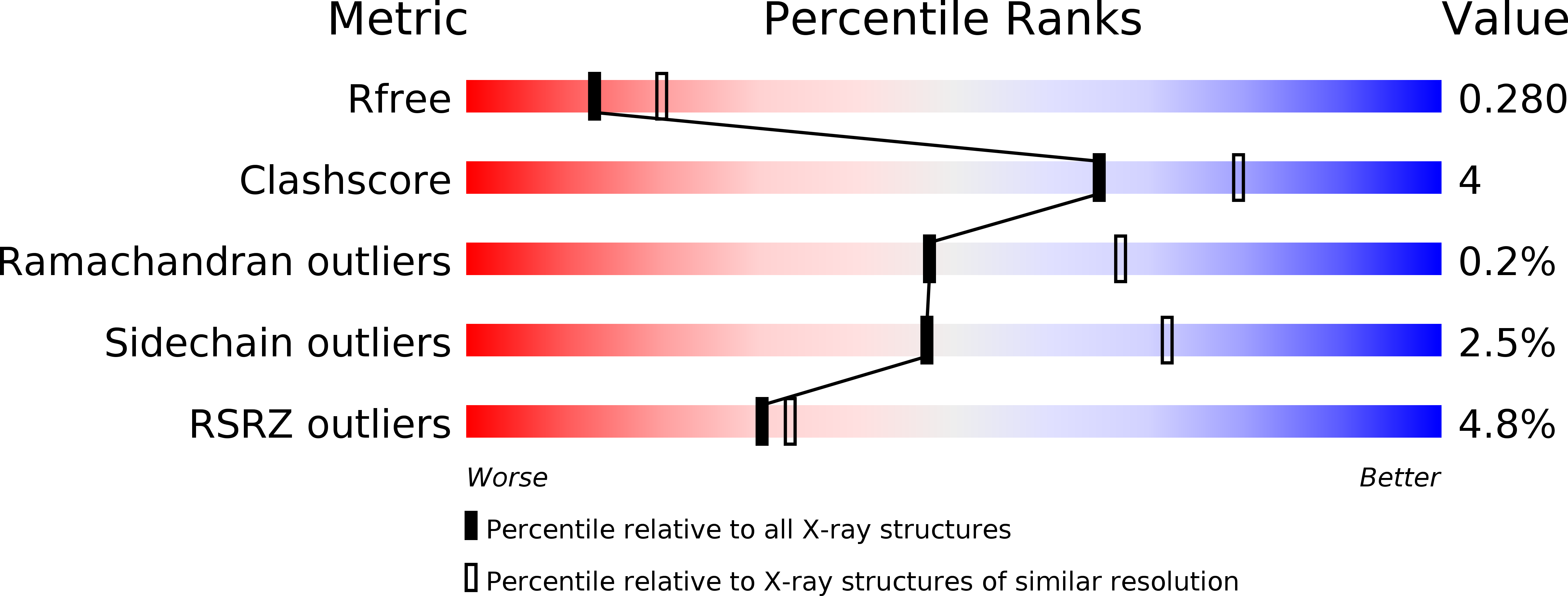
Resolution:
2.53 Å
R-Value Free:
0.28
R-Value Work:
0.24
R-Value Observed:
0.24
Space Group:
P 21 21 21