2VK8 image
Deposition Date 2007-12-17
Release Date 2009-01-27
Last Version Date 2023-12-13
Entry Detail
PDB ID:
2VK8
Keywords:
Title:
Crystal structure of the Saccharomyces cerevisiae pyruvate decarboxylase variant E477Q in complex with its substrate
Biological Source:
Source Organism:
Host Organism:
Method Details:
Experimental Method:
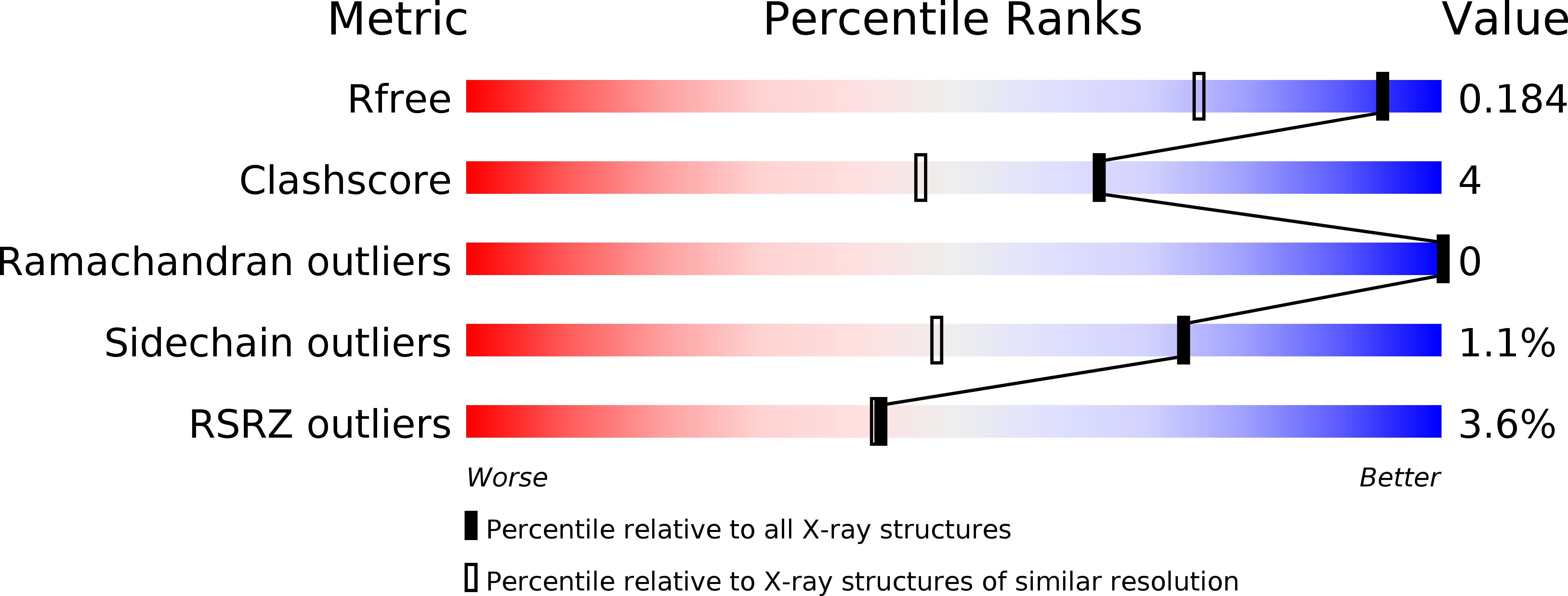
Resolution:
1.42 Å
R-Value Free:
0.18
R-Value Work:
0.18
R-Value Observed:
0.18
Space Group:
P 1 21 1
Macromolecular Entities
Polymer Type:polypeptide(L)
Molecule:PYRUVATE DECARBOXYLASE ISOZYME 1
Gene (Uniprot):PDC1
Mutations:YES
Chain IDs:A, B, C, D
Chain Length:563
Number of Molecules:4
Biological Source:SACCHAROMYCES CEREVISIAE
Primary Citation
Covalently Bound Substrate at the Regulatory Site of Yeast Pyruvate Decarboxylases Triggers Allosteric Enzyme Activation.
J.Biol.Chem. 284 12136 ? (2009)
PMID: 19246454 DOI: 10.1074/JBC.M806228200

Abstact

The mechanism by which the enzyme pyruvate decarboxylase from two yeast species is activated allosterically has been elucidated. A total of seven three-dimensional structures of the enzyme, of enzyme variants, or of enzyme complexes from two yeast species, three of them reported here for the first time, provide detailed atomic resolution snapshots along the activation coordinate. The prime event is the covalent binding of the substrate pyruvate to the side chain of cysteine 221, thus forming a thiohemiketal. This reaction causes the shift of a neighboring amino acid, which eventually leads to the rigidification of two otherwise flexible loops, one of which provides two histidine residues necessary to complete the enzymatically competent active site architecture. The structural data are complemented and supported by kinetic investigations and binding studies, providing a consistent picture of the structural changes occurring upon enzyme activation.

Legend

Protein

Chemical

Disease

Primary Citation of related structures