2VHL image
Entry Detail
PDB ID:
2VHL
Keywords:
Title:
The Three-dimensional structure of the N-Acetylglucosamine-6- phosphate deacetylase from Bacillus subtilis
Biological Source:
Source Organism:
Host Organism:
PDB Version:
Deposition Date:
2007-11-22
Release Date:
2007-12-04
Method Details:
Experimental Method:
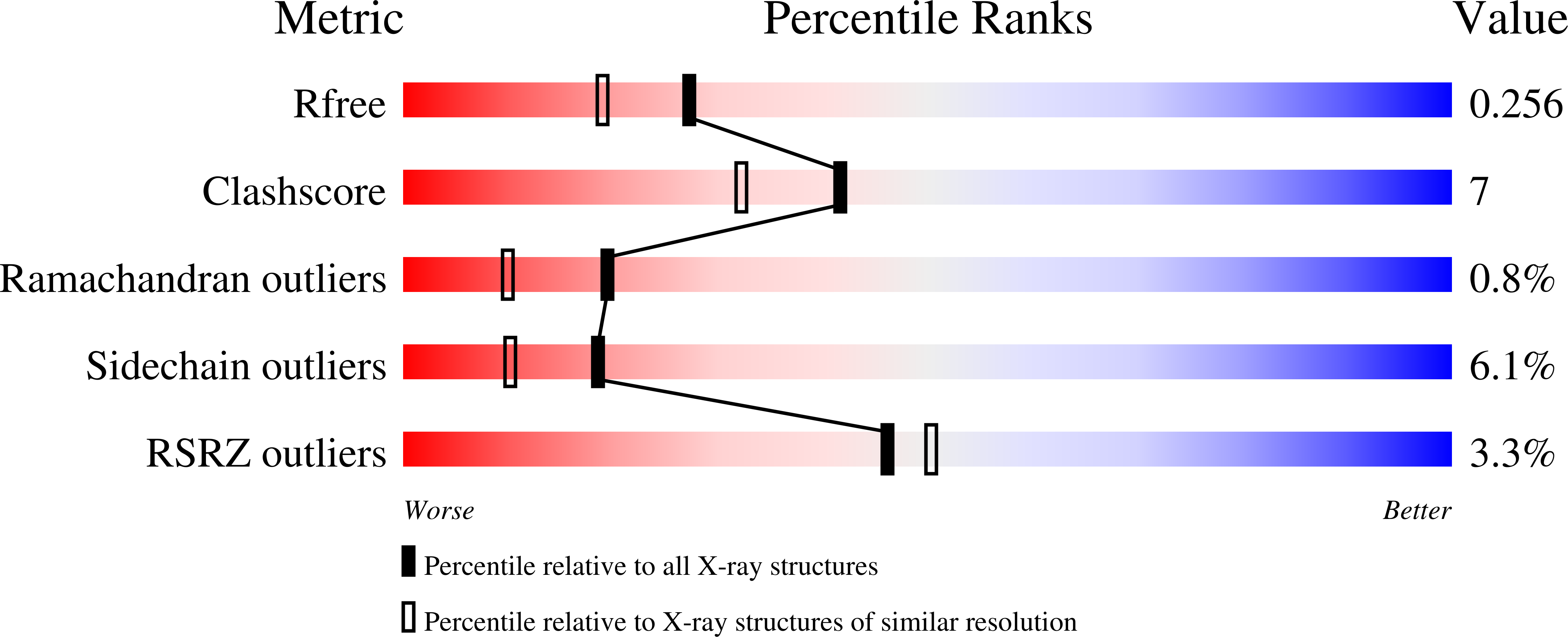
Resolution:
2.05 Å
R-Value Free:
0.26
R-Value Work:
0.20
R-Value Observed:
0.20
Space Group:
P 21 21 2
Macromolecular Entities
Polymer Type:polypeptide(L)
Description:N-ACETYLGLUCOSAMINE-6-PHOSPHATE DEACETYLASE
Chain IDs:A, B
Chain Length:396
Number of Molecules:2
Biological Source:BACILLUS SUBTILIS
Primary Citation
The Three-Dimensional Structure of the N-Acetylglucosamine-6-Phosphate Deacetylase from Bacillus Subtilis
J.Biol.Chem. 279 2809 ? (2004)
PMID: 14557261 DOI: 10.1074/JBC.M310165200

Abstact

The enzyme N-acetylglucosamine-6-phosphate deacetylase, NagA, catalyzes the hydrolysis of the N-acetyl group of GlcNAc-6-P to yield glucosamine 6-phosphate and acetate, the first committed step in the biosynthetic pathway to amino-sugar-nucleotides. It is classified into carbohydrate esterase family CE-9 (see afmb.cnrs-mrs.fr/CAZY/). Here we report the cloning, expression, and three-dimensional structure (Protein Data Bank code 1un7) determination by x-ray crystallography of the Bacillus subtilis NagA at a resolution of 2.0 A. The structure presents two domains, a (beta/alpha)(8) barrel enclosing the active center and a small beta barrel domain. The structure is dimeric, and the substrate phosphate coordination at the active center is provided by an Arg/His pair contributed from the second molecule of the dimer. Both the overall structure and the active center bear a striking similarity to the urease superfamily with two metals involved in substrate binding and catalysis. PIXE (Proton-Induced x-ray Emission) data show that iron is the predominant metal in the purified protein. We propose a catalytic mechanism involving proton donation to the leaving group by aspartate, nucleophilic attack by an Fe-bridged hydroxide, and stabilization of the carbonyl oxygen by one of the two Fe atoms of the pair. We believe that this is the first sugar deacetylase to utilize this fold and catalytic mechanism.

Legend

Protein

Chemical

Disease

Primary Citation of related structures