Deposition Date
2007-11-20
Release Date
2008-11-25
Last Version Date
2024-11-13
Entry Detail
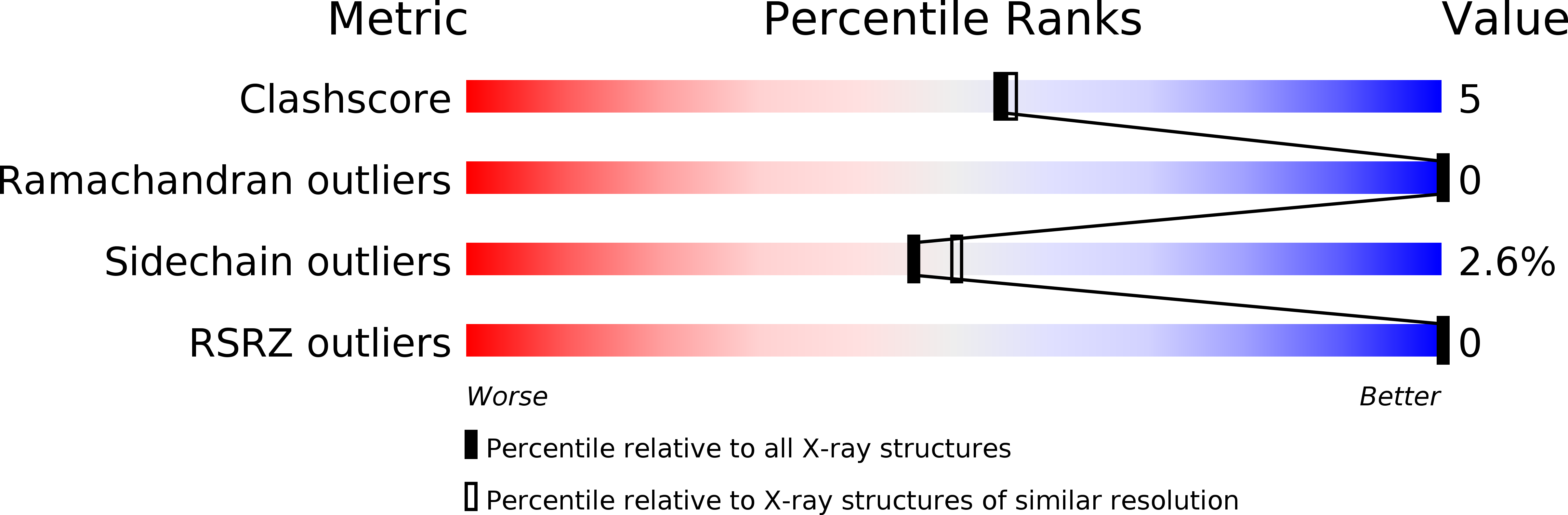
PDB ID:
2VH9
Keywords:
Title:
CRYSTAL STRUCTURE OF NXG1-DELTAYNIIG IN COMPLEX WITH XLLG, A XYLOGLUCAN DERIVED OLIGOSACCHARIDE
Biological Source:
Source Organism(s):
TROPAEOLUM MAJUS (Taxon ID: 4020)
Expression System(s):
Method Details:
Experimental Method:
Resolution:
2.10 Å
R-Value Free:
0.20
R-Value Work:
0.14
R-Value Observed:
0.15
Space Group:
P 31