2VGJ image
Entry Detail
PDB ID:
2VGJ
Keywords:
Title:
Crystal structure of Actinomadura R39 DD-peptidase complexed with a peptidoglycan-mimetic cephalosporin
Biological Source:
Source Organism:
PDB Version:
Deposition Date:
2007-11-14
Release Date:
2008-11-25
Method Details:
Experimental Method:
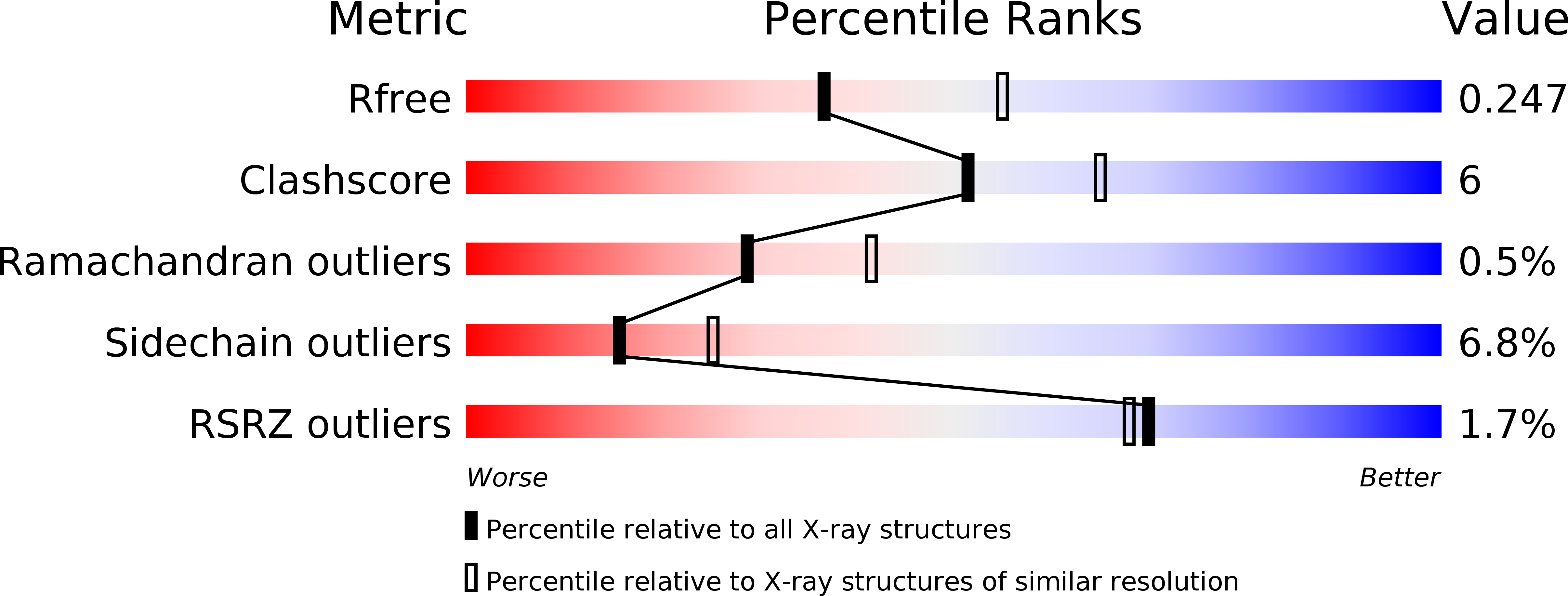
Resolution:
2.40 Å
R-Value Free:
0.24
R-Value Work:
0.19
R-Value Observed:
0.19
Space Group:
P 1 21 1
Macromolecular Entities
Polymer Type:polypeptide(L)
Description:D-ALANYL-D-ALANINE CARBOXYPEPTIDASE
Chain IDs:A, B, C, D
Chain Length:489
Number of Molecules:4
Biological Source:ACTINOMADURA SP.
Primary Citation
Crystal Structures of Complexes of Bacterial Dd-Peptidases with Peptidoglycan-Mimetic Ligands: The Substrate Specificity Puzzle.
J.Mol.Biol. 381 383 ? (2008)
PMID: 18602645 DOI: 10.1016/J.JMB.2008.06.012

Abstact

The X-ray crystal structures of covalent complexes of the Actinomadura R39 dd-peptidase and Escherichia coli penicillin-binding protein (PBP) 5 with beta-lactams bearing peptidoglycan-mimetic side chains have been determined. The structure of the hydrolysis product of an analogous peptide bound noncovalently to the former enzyme has also been obtained. The R39 DD-peptidase structures reveal the presence of a specific binding site for the D-alpha-aminopimelyl side chain, characteristic of the stem peptide of Actinomadura R39. This binding site features a hydrophobic cleft for the pimelyl methylene groups and strong hydrogen bonding to the polar terminus. Both of these active site elements are provided by amino acid side chains from two separate domains of the protein. In contrast, no clear electron density corresponding to the terminus of the peptidoglycan-mimetic side chains is present when these beta-lactams are covalently bound to PBP5. There is, therefore, no indication of a specific side-chain binding site in this enzyme. These results are in agreement with those from kinetics studies published earlier and support the general prediction made at the time of a direct correlation between kinetics and structural evidence. The essential high-molecular-mass PBPs have demonstrated, to date, no specific reactivity with peptidoglycan-mimetic peptide substrates and beta-lactam inhibitors and, thus, probably do not possess a specific substrate-binding site of the type demonstrated here with the R39 DD-peptidase. This striking deficiency may represent a sophisticated defense mechanism against low-molecular-mass substrate-analogue inhibitors/antibiotics; its discovery should focus new inhibitor design.

Legend

Protein

Chemical

Disease

Primary Citation of related structures