
Deposition Date
2007-10-31
Release Date
2008-12-16
Last Version Date
2023-12-13
Entry Detail
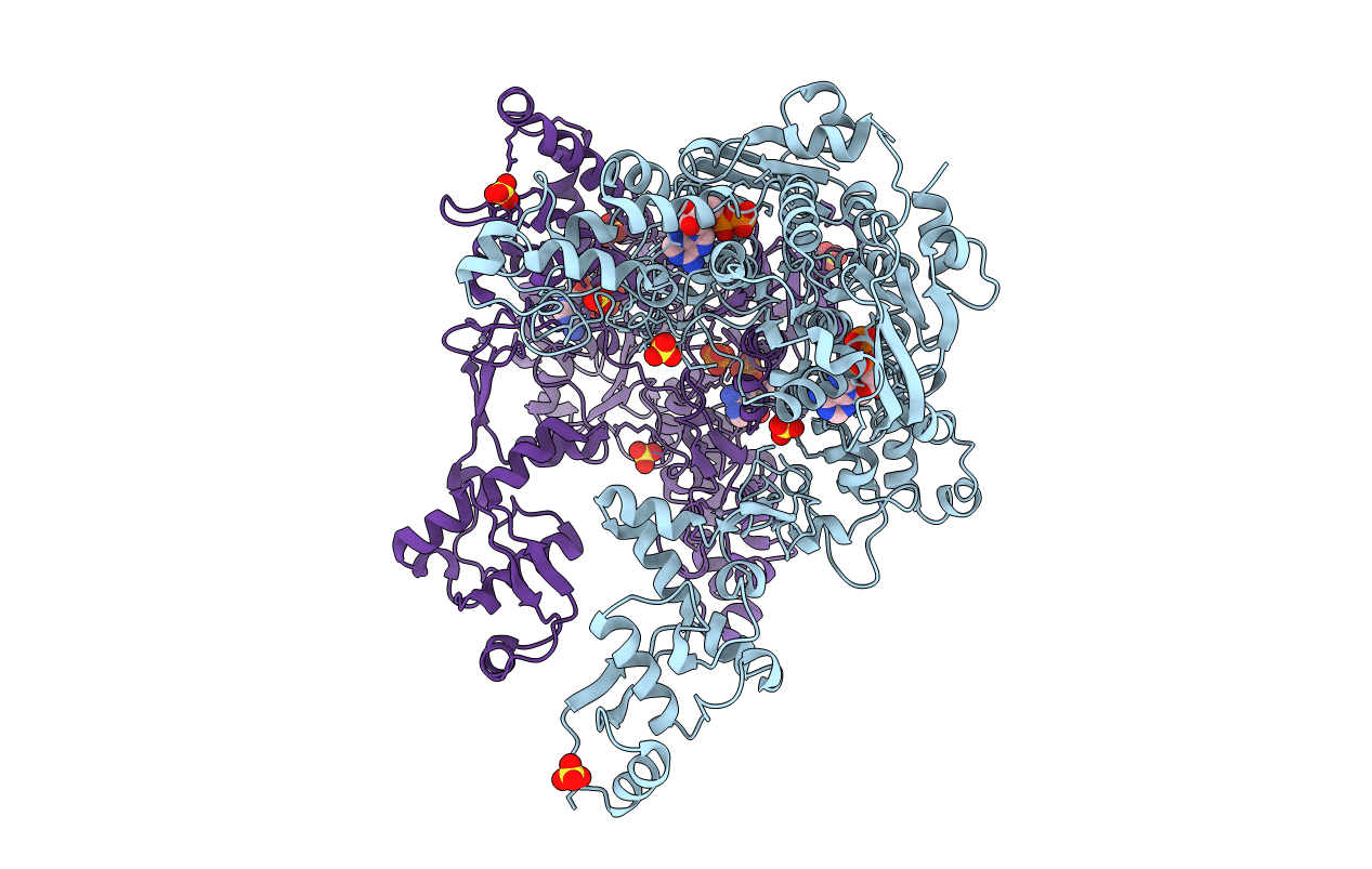
PDB ID:
2VF8
Keywords:
Title:
Crystal structure of UvrA2 from Deinococcus radiodurans
Biological Source:
Source Organism(s):
DEINOCOCCUS RADIODURANS (Taxon ID: 1299)
Expression System(s):
Method Details:
Experimental Method:
Resolution:
3.00 Å
R-Value Free:
0.29
R-Value Work:
0.21
R-Value Observed:
0.22
Space Group:
C 2 2 21
