2VEP image
Deposition Date 2007-10-25
Release Date 2007-11-20
Last Version Date 2024-05-08
Entry Detail
PDB ID:
2VEP
Keywords:
Title:
Crystal Structure Of The Full Length Bifunctional Enzyme Pria
Biological Source:
Source Organism:
Method Details:
Experimental Method:
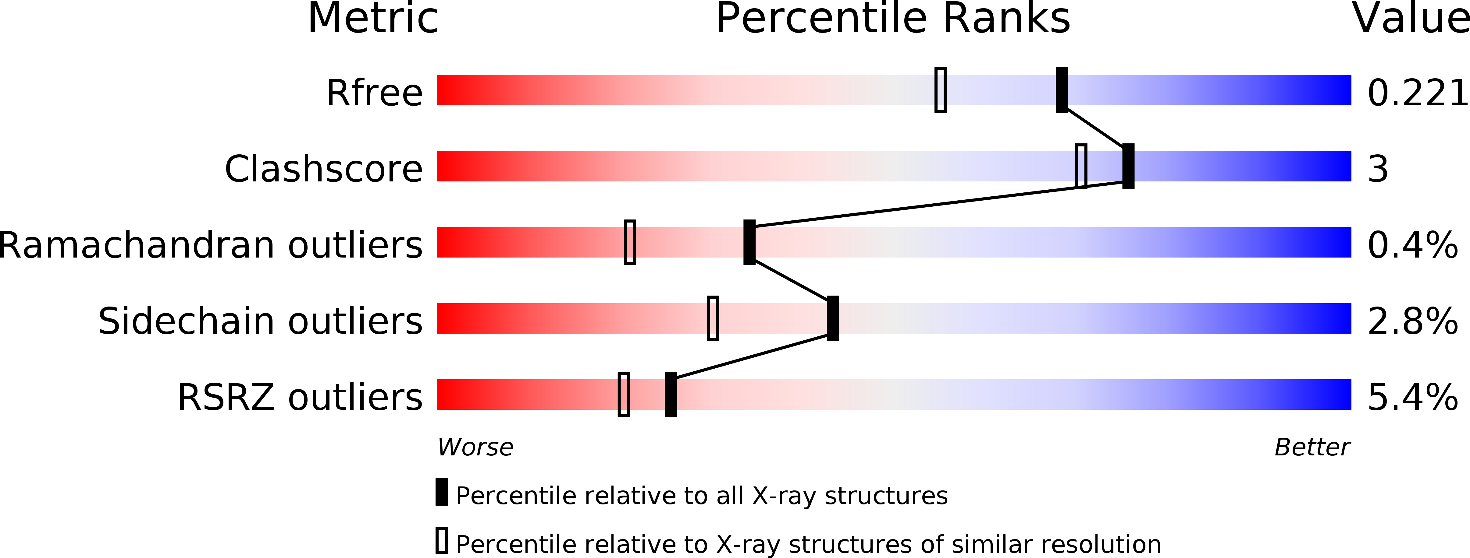
Resolution:
1.80 Å
R-Value Free:
0.22
R-Value Work:
0.18
R-Value Observed:
0.18
Space Group:
P 31 2 1
Macromolecular Entities
Structures with similar UniProt ID
Protein Blast
Polymer Type:polypeptide(L)
Molecule:PHOSPHORIBOSYL ISOMERASE A
Gene (Uniprot):priA
Chain IDs:A
Chain Length:244
Number of Molecules:1
Biological Source:STREPTOMYCES COELICOLOR
Ligand Molecules
Primary Citation
The Structure/Function Relationship of a Dual Substrate (Betaalpha)(8)-Isomerase
Biochem.Biophys.Res.Commun. 365 16 ? (2008)
PMID: 17967415 DOI: 10.1016/J.BBRC.2007.10.101

Abstact

Two structures of phosphoribosyl isomerase A (PriA) from Streptomyces coelicolor, involved in both histidine and tryptophan biosynthesis, were solved at 1.8A resolution. A closed conformer was obtained, which represents the first complete structure of PriA, revealing hitherto unnoticed molecular interactions and the occurrence of conformational changes. Inspection of these conformers, including ligand-docking simulations, allowed identification of residues involved in substrate recognition, chemical catalysis and conformational changes. These predictions were validated by mutagenesis and functional analysis. Arg19 and Ser81 were shown to play critical roles within the carboxyl and amino phosphate-binding sites, respectively; the catalytic residues Asp11 and Asp130 are responsible for both activities; and Thr166 and Asp171, which make an unusual contact, are likely to elicit the conformational changes needed for adopting the active site architectures. This represents the first report of the structure/function relationship of this (betaalpha)8-isomerase.

Legend

Protein

Chemical

Disease

Primary Citation of related structures
Feedback Form
Name
Email
Institute
Feedback