2VEE image
Entry Detail
PDB ID:
2VEE
Title:
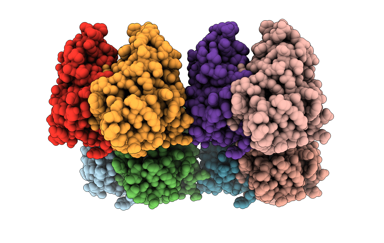
Structure of protoglobin from Methanosarcina acetivorans C2A
Biological Source:
Host Organism:
PDB Version:
Deposition Date:
2007-10-22
Release Date:
2008-01-22
Method Details:
Experimental Method:
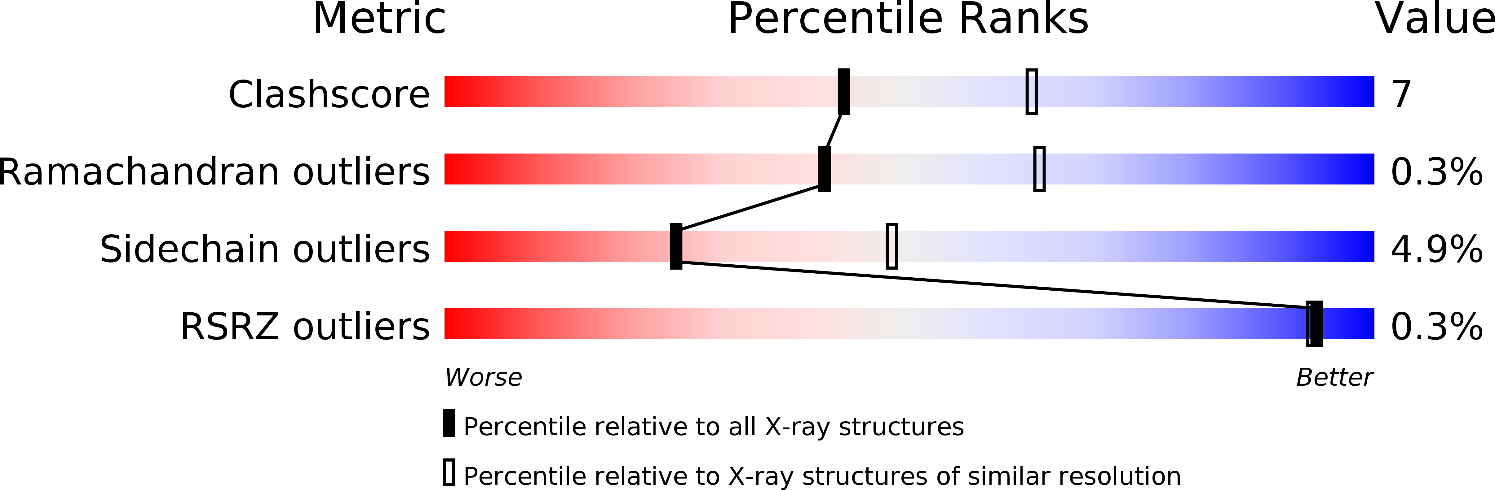
Resolution:
2.60 Å
R-Value Free:
0.26
R-Value Work:
0.20
R-Value Observed:
0.20
Space Group:
P 1 21 1
Macromolecular Entities
Polymer Type:polypeptide(L)
Description:PROTOGLOBIN
Mutations:YES
Chain IDs:A, B, C, D, E, F, G, H
Chain Length:195
Number of Molecules:8
Biological Source:METHANOSARCINA ACETIVORANS
Ligand Molecules
Primary Citation
Archaeal Protoglobin Structure Indicates New Ligand Diffusion Paths and Modulation of Haem-Reactivity.
Embo Rep. 9 157 ? (2008)
PMID: 18188182 DOI: 10.1038/SJ.EMBOR.7401153

Abstact

The structural adaptability of the globin fold has been highlighted by the recent discovery of the 2-on-2 haemoglobins, of neuroglobin and cytoglobin. Protoglobin from Methanosarcina acetivorans C2A-a strictly anaerobic methanogenic Archaea-is, to the best of our knowledge, the latest entry adding new variability and functional complexity to the haemoglobin (Hb) superfamily. Here, we report the 1.3 A crystal structure of oxygenated M. acetivorans protoglobin, together with the first insight into its ligand-binding properties. We show that, contrary to all known globins, protoglobin-specific loops and an amino-terminal extension completely bury the haem within the protein matrix. Access of O(2), CO and NO to the haem is granted by the protoglobin-specific apolar tunnels reaching the haem distal site from locations at the B/G and B/E helix interfaces. Functionally, M. acetivorans dimeric protoglobin shows a selectivity ratio for O(2)/CO binding to the haem that favours O(2) ligation and anticooperativity in ligand binding. Both properties are exceptional within the Hb superfamily.

Legend

Protein

Chemical

Disease

Primary Citation of related structures