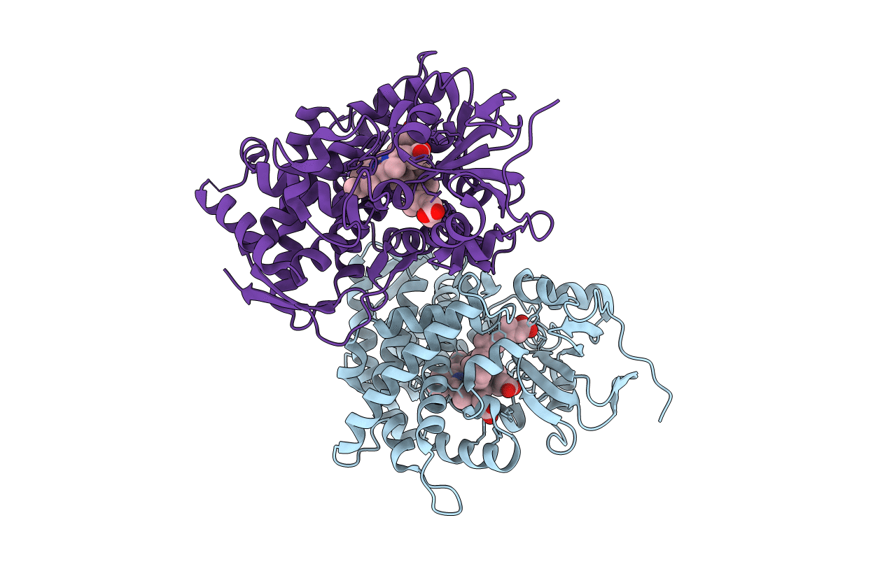
2VE3 image
Entry Detail
PDB ID:
2VE3
Keywords:
Title:
Retinoic acid bound cyanobacterial CYP120A1
Biological Source:
Source Organism:
Host Organism:
PDB Version:
Deposition Date:
2007-10-15
Release Date:
2008-04-29
Method Details:
Experimental Method:
Resolution:
2.10 Å
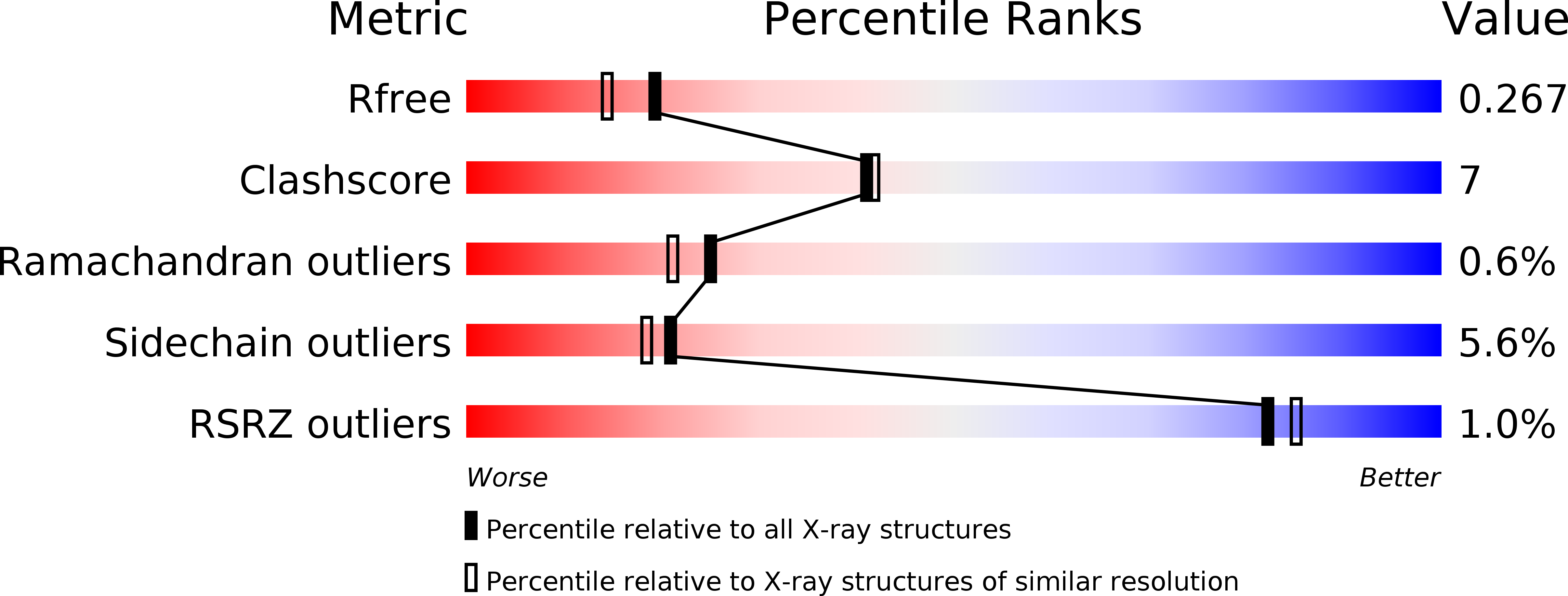
R-Value Free:
0.26
R-Value Work:
0.22
R-Value Observed:
0.22
Space Group:
C 1 2 1
Macromolecular Entities
Polymer Type:polypeptide(L)
Description:PUTATIVE CYTOCHROME P450 120
Chain IDs:A, B
Chain Length:444
Number of Molecules:2
Biological Source:SYNECHOCYSTIS SP.
Primary Citation
Crystal Structures of Substrate-Free and Retinoic Acid-Bound Cyanobacterial Cytochrome P450 Cyp120A1.
Biochemistry 47 6552 ? (2008)
PMID: 18512957 DOI: 10.1021/BI800328S

Abstact

The crystal structures of substrate-free and all-trans-retinoic acid-bound CYP120A1 from Synechocystis sp. PCC 6803 were determined at 2.4 and 2.1 A resolution, respectively, representing the first structural characterization of a cyanobacterial P450. Features of CYP120A1 not observed in other P450 structures include an aromatic ladder flanking the channel leading to the active site and a triple-glycine motif within SRS5. Using spectroscopic methods, CYP120A1 is shown to bind 13-cis-retinoic acid, 9-cis-retinoic acid, and retinal with high affinity and dissociation constants of less than 1 microM. Metabolism of retinoic acid by CYP120A1 suggests that CYP120A1 hydroxylates a variety of retinoid derivatives in vivo. On the basis of the retinoic acid-bound CYP120A1 crystal structure, we propose that either carbon 2 or the methyl groups (C16 or C17) of the beta-ionone ring are modified by CYP120A1.

Legend

Protein

Chemical

Disease

Primary Citation of related structures