
Deposition Date
2007-10-10
Release Date
2008-09-02
Last Version Date
2024-11-13
Entry Detail
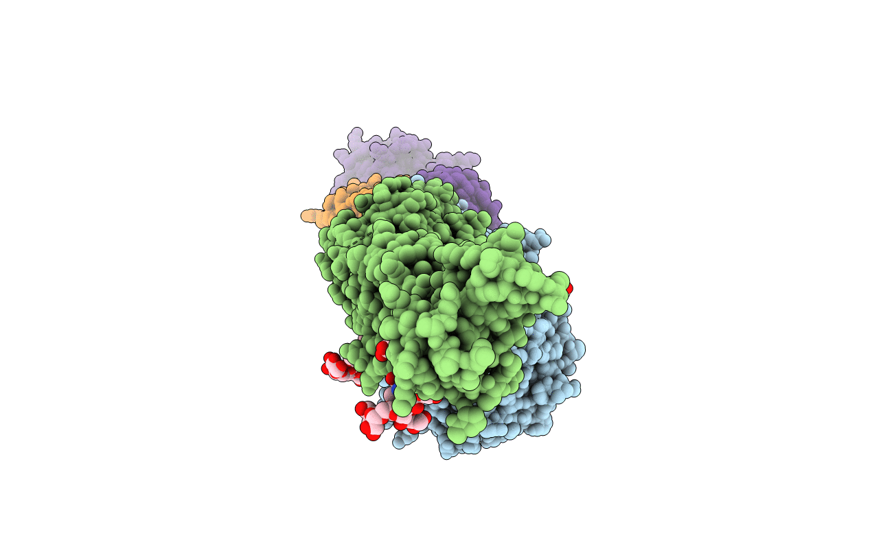
PDB ID:
2VDR
Keywords:
Title:
Integrin AlphaIIbBeta3 Headpiece Bound to a chimeric Fibrinogen Gamma chain peptide, LGGAKQRGDV
Biological Source:
Source Organism(s):
HOMO SAPIENS (Taxon ID: 9606)
MUS MUSCULUS (Taxon ID: 10090)
MUS MUSCULUS (Taxon ID: 10090)
Expression System(s):
Method Details:
Experimental Method:
Resolution:
2.40 Å
R-Value Free:
0.19
R-Value Work:
0.14
R-Value Observed:
0.15
Space Group:
P 32 2 1
