2VD5 image
Entry Detail
PDB ID:
2VD5
Keywords:
Title:
Structure of Human Myotonic Dystrophy Protein Kinase in Complex with the Bisindoylmaleide inhibitor BIM VIII
Biological Source:
Source Organism:
Host Organism:
PDB Version:
Deposition Date:
2007-09-30
Release Date:
2007-11-06
Method Details:
Experimental Method:
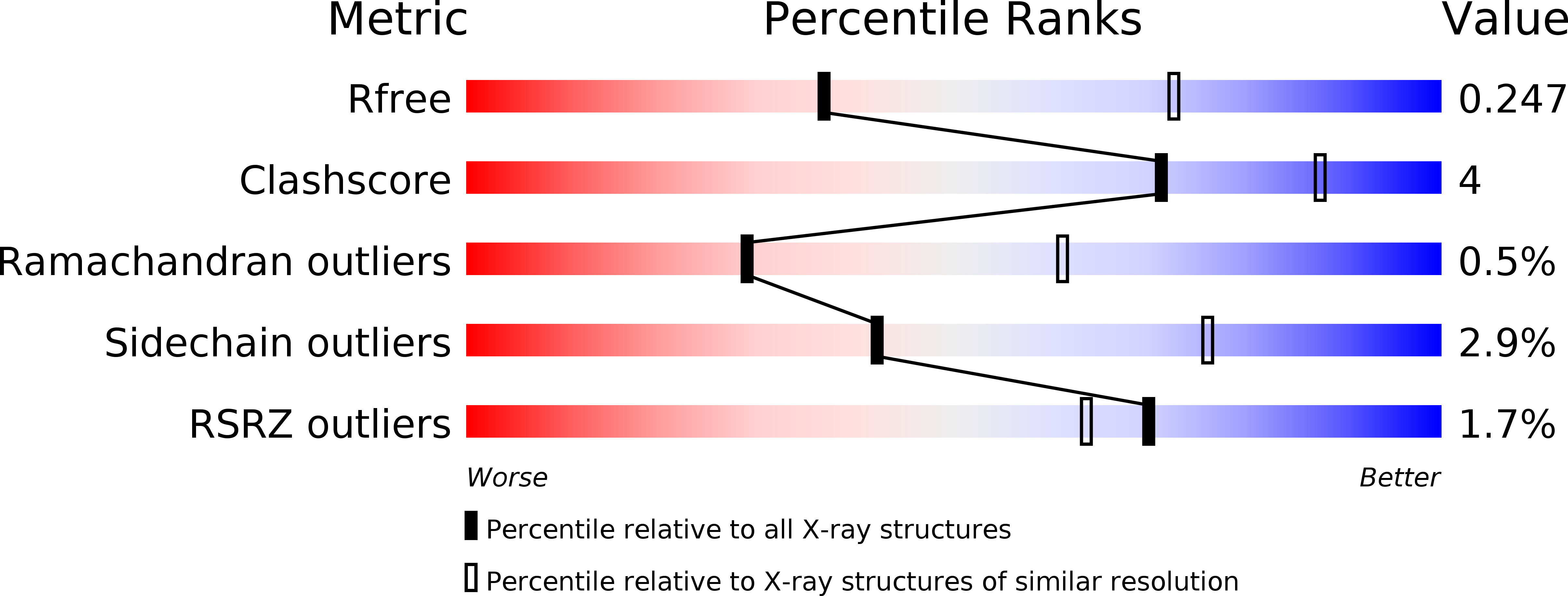
Resolution:
2.80 Å
R-Value Free:
0.24
R-Value Work:
0.19
R-Value Observed:
0.19
Space Group:
P 32 2 1
Macromolecular Entities
Polymer Type:polypeptide(L)
Description:DMPK PROTEIN
Chain IDs:A, B
Chain Length:412
Number of Molecules:2
Biological Source:HOMO SAPIENS
Ligand Molecules
Primary Citation
Structure of Dystrophia Myotonica Protein Kinase.
Protein Sci. 18 782 ? (2009)
PMID: 19309729 DOI: 10.1002/PRO.82

Abstact

Dystrophia myotonica protein kinase (DMPK) is a serine/threonine kinase composed of a kinase domain and a coiled-coil domain involved in the multimerization. The crystal structure of the kinase domain of DMPK bound to the inhibitor bisindolylmaleimide VIII (BIM-8) revealed a dimeric enzyme associated by a conserved dimerization domain. The affinity of dimerisation suggested that the kinase domain alone is insufficient for dimerisation in vivo and that the coiled-coil domains are required for stable dimer formation. The kinase domain is in an active conformation, with a fully-ordered and correctly positioned alphaC helix, and catalytic residues in a conformation competent for catalysis. The conserved hydrophobic motif at the C-terminal extension of the kinase domain is bound to the N-terminal lobe of the kinase domain, despite being unphosphorylated. Differences in the arrangement of the C-terminal extension compared to the closely related Rho-associated kinases include an altered PXXP motif, a different conformation and binding arrangement for the turn motif, and a different location for the conserved NFD motif. The BIM-8 inhibitor occupies the ATP site and has similar binding mode as observed in PDK1.

Legend

Protein

Chemical

Disease

Primary Citation of related structures