2VCP image
Deposition Date 2007-09-26
Release Date 2008-11-04
Last Version Date 2023-12-13
Entry Detail
PDB ID:
2VCP
Title:
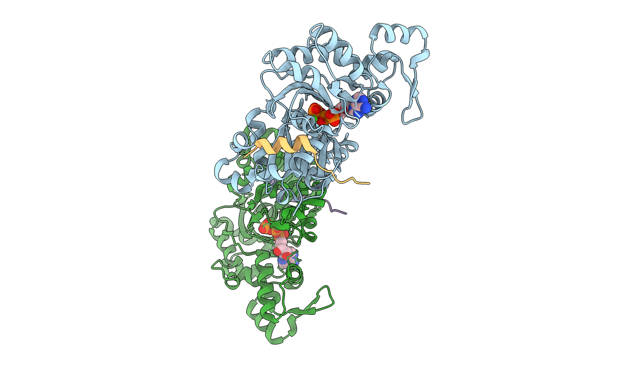
Crystal structure of N-Wasp VC domain in complex with skeletal actin
Biological Source:
Source Organism:
Host Organism:
Method Details:
Experimental Method:
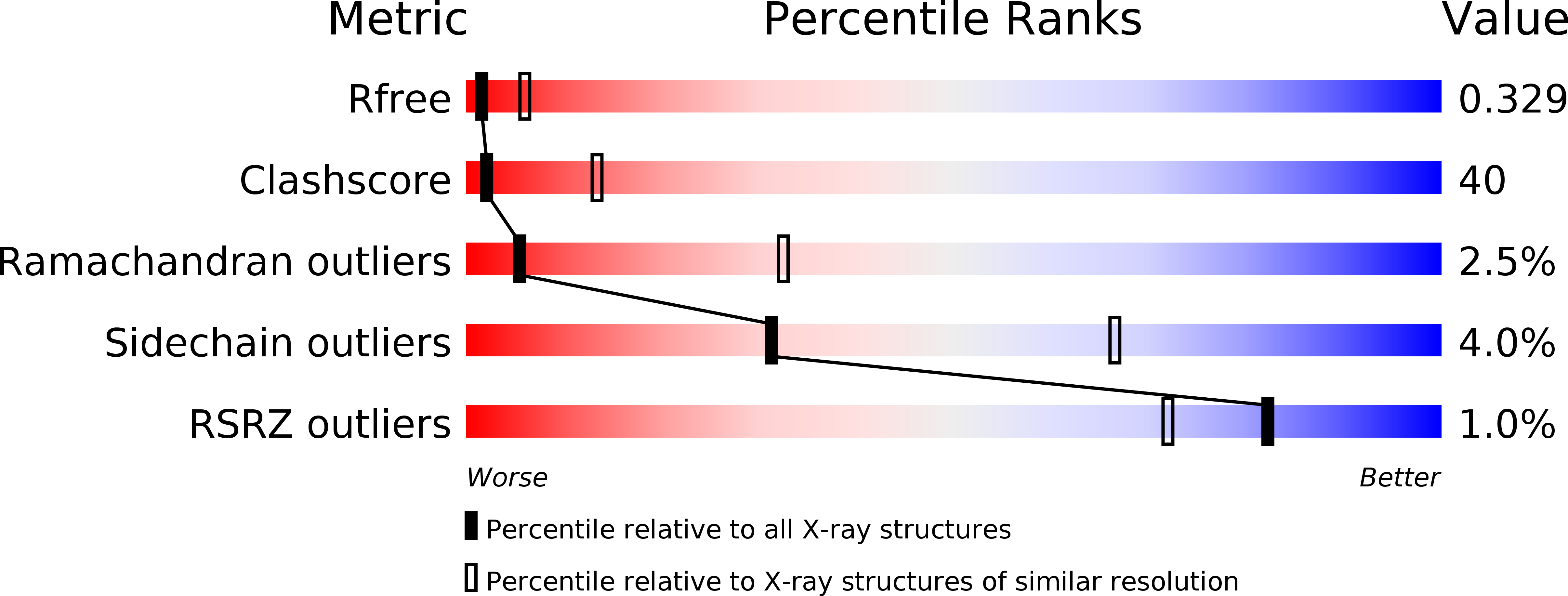
Resolution:
3.20 Å
R-Value Free:
0.33
R-Value Work:
0.27
R-Value Observed:
0.27
Space Group:
P 61
Macromolecular Entities
Structures with similar UniProt ID
Protein Blast
Polymer Type:polypeptide(L)
Molecule:ACTIN, ALPHA SKELETAL MUSCLE
Gene (Uniprot):ACTA1
Chain IDs:A, B
Chain Length:375
Number of Molecules:2
Biological Source:ORYCTOLAGUS CUNICULUS
Structures with similar UniProt ID
Protein Blast
Polymer Type:polypeptide(L)
Molecule:NEURAL WISKOTT-ALDRICH SYNDROME PROTEIN
Gene (Uniprot):WASL
Chain IDs:C (auth: D), D (auth: E)
Chain Length:93
Number of Molecules:2
Biological Source:HOMO SAPIENS
Modified Residue
Compound ID Chain ID Parent Comp ID Details 2D Image
HIC A HIS 4-METHYL-HISTIDINE
Primary Citation
Interactions of isolated C-terminal fragments of neural Wiskott-Aldrich syndrome protein (N-WASP) with actin and Arp2/3 complex.
J. Biol. Chem. 287 34646 34659 (2012)
PMID: 22847007 DOI: 10.1074/jbc.M112.394361

Abstact

Wiskott-Aldrich syndrome proteins (WASP) are a family of proteins that all catalyze actin filament branching with the Arp2/3 complex in a variety of actin-based motile processes. The constitutively active C-terminal domain, called VCA, harbors one or more WASP homology 2 (WH2) domains that bind G-actin, whereas the CA extension binds the Arp2/3 complex. The VCA·actin·Arp2/3 entity associates with a mother filament to form a branched junction from which a daughter filament is initiated. The number and function of WH2-bound actin(s) in the branching process are not known, and the stoichiometry of the VCA·actin·Arp2/3 complex is debated. We have expressed the tandem WH2 repeats of N-WASP, either alone (V) or associated with the C (VC) and CA (VCA) extensions. We analyzed the structure of actin in complex with V, VC, and VCA using protein crystallography and hydrodynamic and spectrofluorimetric methods. The partial crystal structure of the VC·actin 1:1 complex shows two actins in the asymmetric unit with extensive actin-actin contacts. In solution, each of the two WH2 domains in V, VC, and VCA binds G-actin in 1:2 complexes that participate in barbed end assembly. V, VC, and VCA enhance barbed end depolymerization like profilin but neither nucleate nor sever filaments, in contrast with other WH2 repeats. VCA binds the Arp2/3 complex in a 1:1 complex even in the presence of a large excess of VCA. VCA·Arp2/3 binds one actin in a latrunculin A-sensitive fashion, in a 1:1:1 complex, indicating that binding of the second actin to VCA is weakened in the ternary complex.

Legend

Protein

Chemical

Disease

Feedback Form
Name
Email
Institute
Feedback