2VAJ image
Entry Detail
PDB ID:
2VAJ
Keywords:
Title:
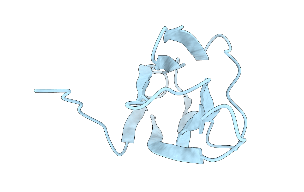
Crystal structure of NCAM2 Ig1 (I4122 cell unit)
Biological Source:
Source Organism:
PDB Version:
Deposition Date:
2007-08-31
Release Date:
2008-08-26
Method Details:
Experimental Method:
Resolution:
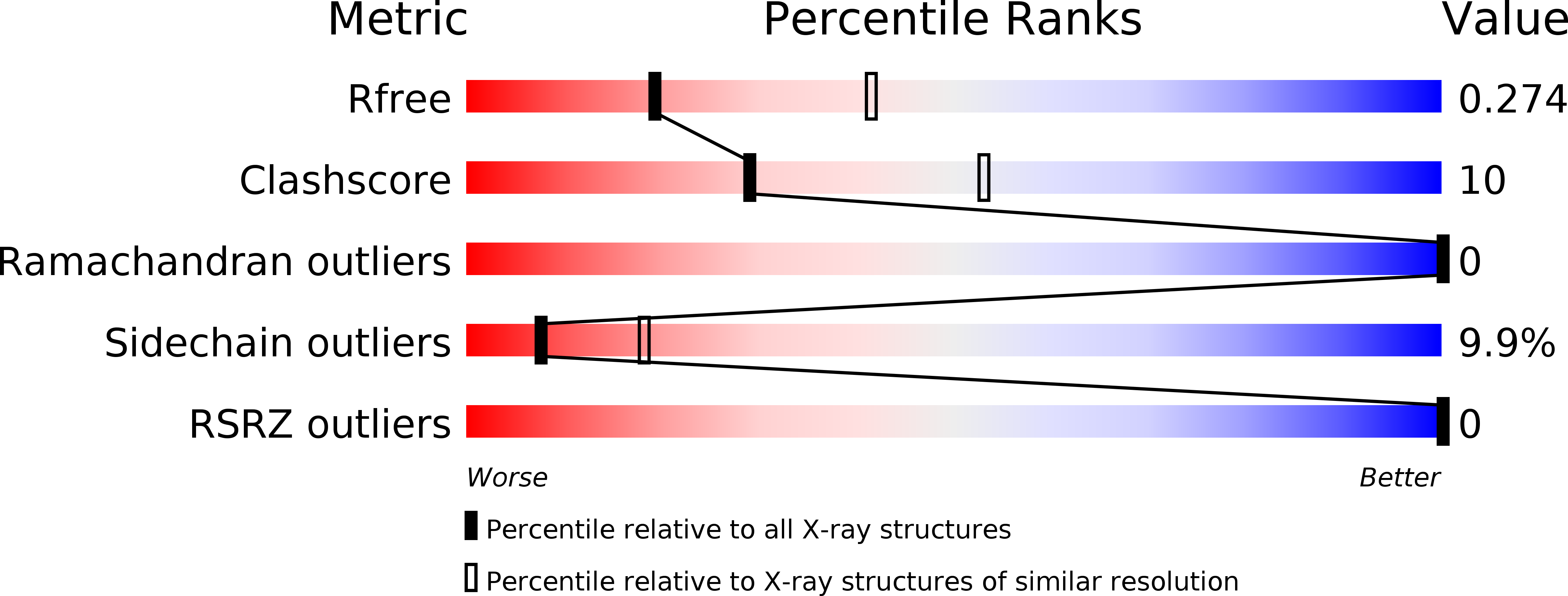
2.70 Å
R-Value Free:
0.26
R-Value Work:
0.22
R-Value Observed:
0.22
Space Group:
I 41 2 2
Macromolecular Entities
Polymer Type:polypeptide(L)
Description:NEURAL CELL ADHESION MOLECULE 2
Chain IDs:A
Chain Length:93
Number of Molecules:1
Biological Source:HOMO SAPIENS
Primary Citation
Crystal Structure of the Ig1 Domain of the Neural Cell Adhesion Molecule Ncam2 Displays Domain Swapping.
J.Mol.Biol. 382 1113 ? (2008)
PMID: 18706912 DOI: 10.1016/J.JMB.2008.07.084

Abstact

The crystal structure of the first immunoglobulin (Ig1) domain of neural cell adhesion molecule 2 (NCAM2/OCAM/RNCAM) is presented at a resolution of 2.7 A. NCAM2 is a member of the immunoglobulin superfamily of cell adhesion molecules (IgCAMs). In the structure, two Ig domains interact by domain swapping, as the two N-terminal beta-strands are interchanged. beta-Strand swapping at the terminal domain is the accepted mechanism of homophilic interactions amongst the cadherins, another class of CAMs, but it has not been observed within the IgCAM superfamily. Gel-filtration chromatography demonstrated the ability of NCAM2 Ig1 to form dimers in solution. Taken together, these observations suggest that beta-strand swapping could have a role in the molecular mechanism of homophilic binding for NCAM2.

Legend

Protein

Chemical

Disease

Primary Citation of related structures