Deposition Date
2007-08-16
Release Date
2007-08-28
Last Version Date
2023-12-13
Entry Detail
PDB ID:
2V8Y
Keywords:
Title:
Crystallographic and mass spectrometric characterisation of eIF4E with N7-cap derivatives
Biological Source:
Source Organism(s):
HOMO SAPIENS (Taxon ID: 9606)
Expression System(s):
Method Details:
Experimental Method:
Resolution:
2.10 Å
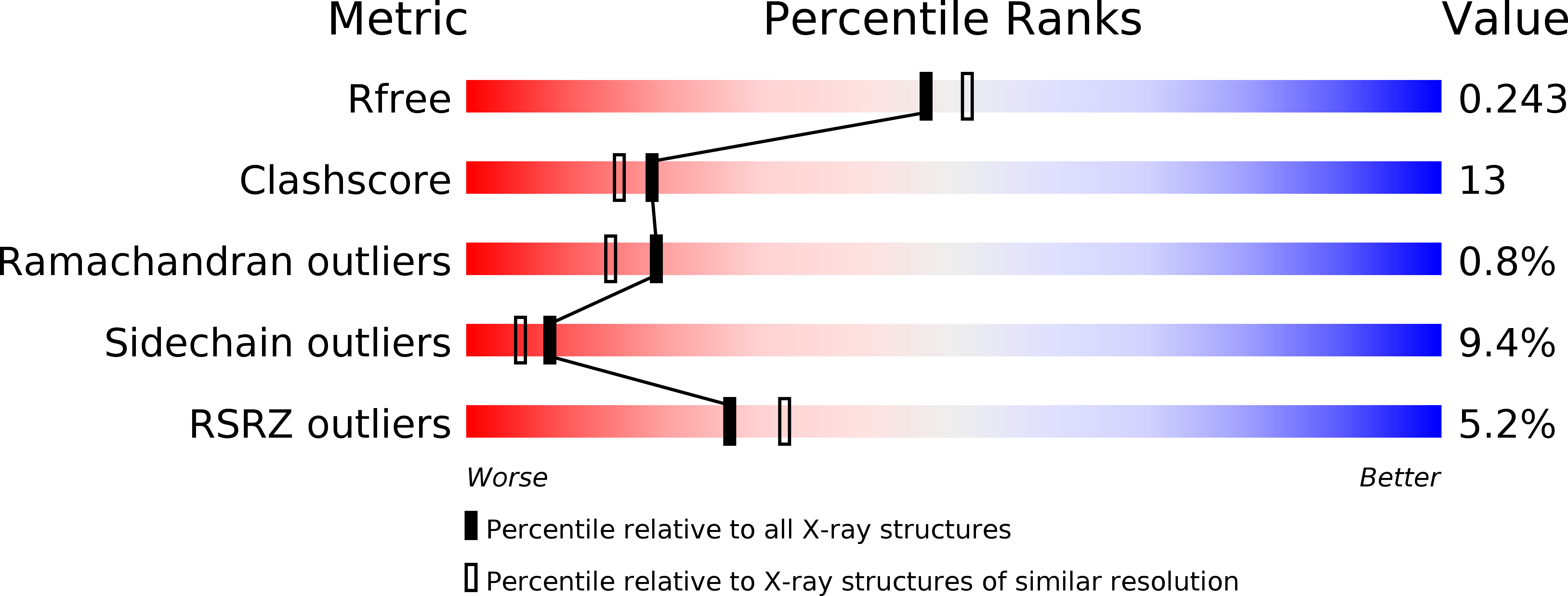
R-Value Free:
0.25
R-Value Work:
0.20
R-Value Observed:
0.20
Space Group:
P 21 21 21