2V8L image
Entry Detail
PDB ID:
2V8L
Keywords:
Title:
Carbohydrate-binding of the starch binding domain of Rhizopus oryzae glucoamylase in complex with beta-cyclodextrin and maltoheptaose
Biological Source:
Source Organism:
Host Organism:
PDB Version:
Deposition Date:
2007-08-09
Release Date:
2008-08-19
Method Details:
Experimental Method:
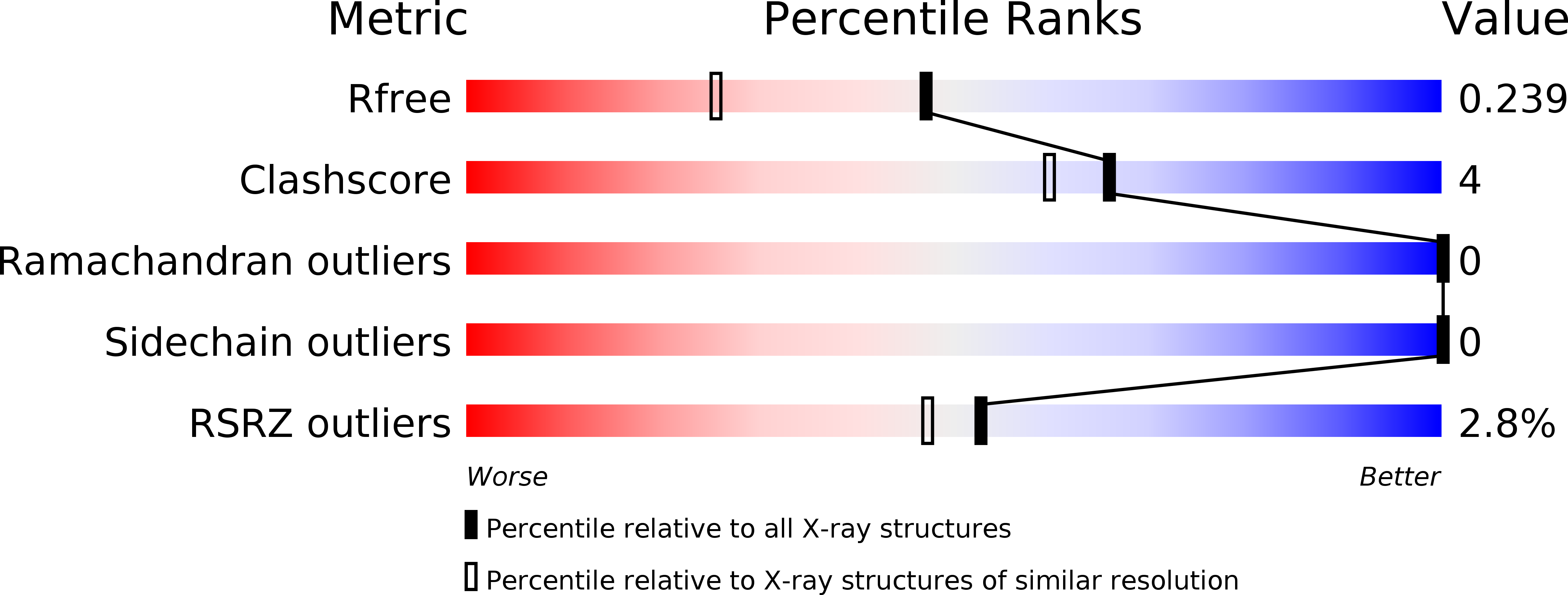
Resolution:
1.80 Å
R-Value Free:
0.23
R-Value Work:
0.22
R-Value Observed:
0.22
Space Group:
P 21 21 21
Macromolecular Entities
Polymer Type:polypeptide(L)
Description:GLUCOAMYLASE A
Chain IDs:A
Chain Length:106
Number of Molecules:1
Biological Source:RHIZOPUS ORYZAE
Ligand Molecules
Peptide-like Molecules
PRD_900012
Primary Citation
Crystal Structures of the Starch-Binding Domain from Rhizopus Oryzae Glucoamylase Reveal a Polysaccharide-Binding Path.
Biochem.J. 416 27 ? (2008)
PMID: 18588504 DOI: 10.1042/BJ20080580

Abstact

GA (glucoamylase) hydrolyses starch and polysaccharides to beta-D-glucose. RoGA (Rhizopus oryzae GA) consists of two functional domains, an N-terminal SBD (starch-binding domain) and a C-terminal catalytic domain, which are connected by an O-glycosylated linker. In the present study, the crystal structures of the SBD from RoGA (RoGACBM21) and the complexes with beta-cyclodextrin (SBD-betaCD) and maltoheptaose (SBD-G7) were determined. Two carbohydrate binding sites, I (Trp(47)) and II (Tyr(32)), were resolved and their binding was co-operative. Besides the hydrophobic interaction, two unique polyN loops comprising consecutive asparagine residues also participate in the sugar binding. A conformational change in Tyr(32) was observed between unliganded and liganded SBDs. To elucidate the mechanism of polysaccharide binding, a number of mutants were constructed and characterized by a quantitative binding isotherm and Scatchard analysis. A possible binding path for long-chain polysaccharides in RoGACBM21 was proposed.

Legend

Protein

Chemical

Disease

Primary Citation of related structures