Deposition Date
2007-07-31
Release Date
2007-09-11
Last Version Date
2024-11-13
Entry Detail
PDB ID:
2V7O
Keywords:
Title:
Crystal structure of human calcium-calmodulin-dependent protein kinase II gamma
Biological Source:
Source Organism(s):
HOMO SAPIENS (Taxon ID: 9606)
Expression System(s):
Method Details:
Experimental Method:
Resolution:
2.25 Å
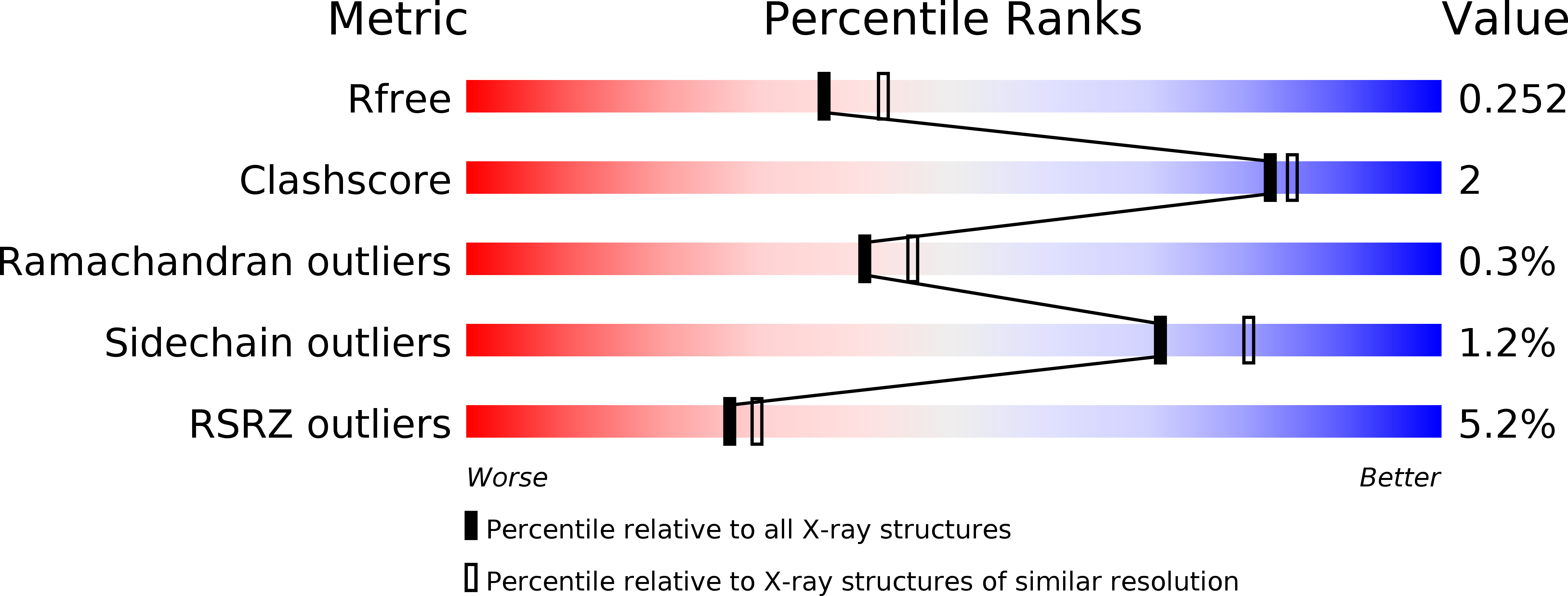
R-Value Free:
0.24
R-Value Work:
0.17
R-Value Observed:
0.17
Space Group:
C 2 2 21