2V6H image
Deposition Date 2007-07-18
Release Date 2008-07-22
Last Version Date 2024-05-08
Entry Detail
PDB ID:
2V6H
Keywords:
Title:
Crystal structure of the C1 domain of cardiac myosin binding protein-C
Biological Source:
Source Organism(s):
HOMO SAPIENS (Taxon ID: 9606)
Expression System(s):
Method Details:
Experimental Method:
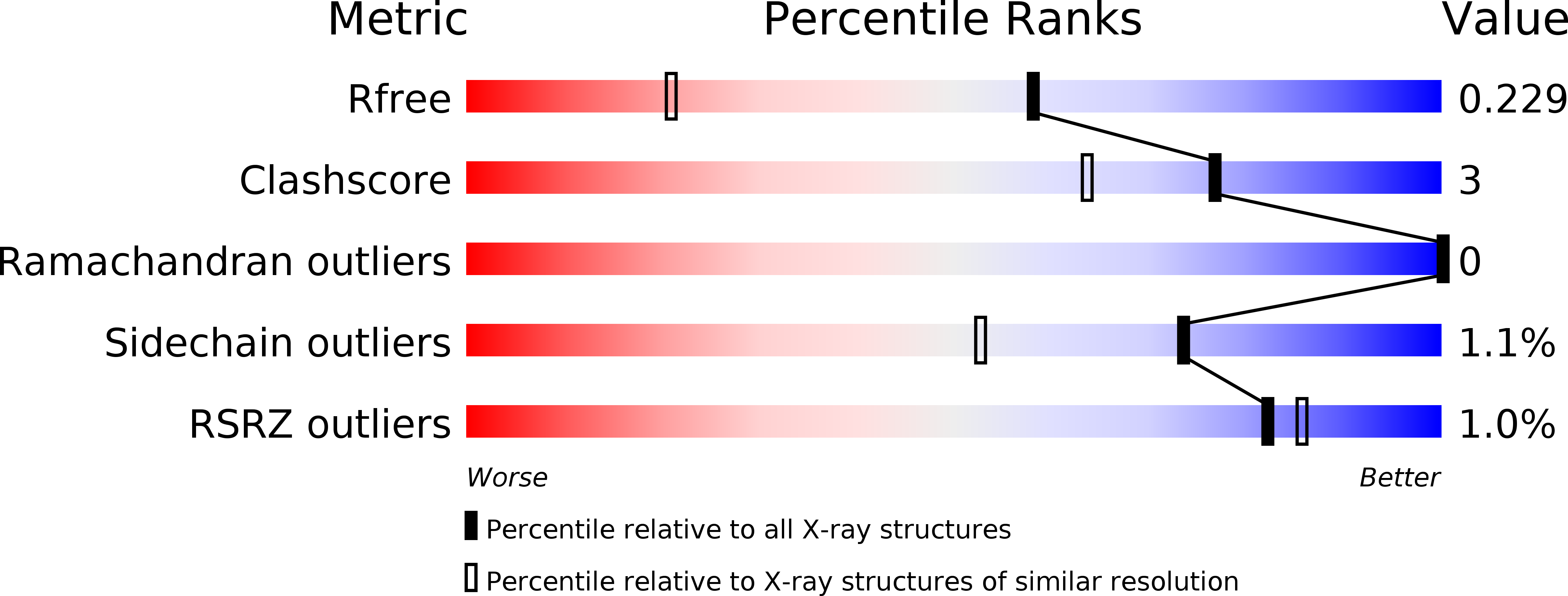
Resolution:
1.55 Å
R-Value Free:
0.24
R-Value Observed:
0.18
Space Group:
I 41
Macromolecular Entities
Structures with similar UniProt ID
Protein Blast
Polymer Type:polypeptide(L)
Molecule:MYOSIN-BINDING PROTEIN C, CARDIAC-TYPE
Gene (Uniprot):MYBPC3
Chain IDs:A
Chain Length:108
Number of Molecules:1
Biological Source:HOMO SAPIENS
Primary Citation
Crystal structure of the C1 domain of cardiac myosin binding protein-C: implications for hypertrophic cardiomyopathy.
J. Mol. Biol. 378 387 397 (2008)
PMID: 18374358 DOI: 10.1016/j.jmb.2008.02.044

Abstact

C-protein is a major component of skeletal and cardiac muscle thick filaments. Mutations in the gene encoding cardiac C-protein [cardiac myosin binding protein-C (cMyBP-C)] are one of the principal causes of hypertrophic cardiomyopathy. cMyBP-C is a string of globular domains including eight immunoglobulin-like and three fibronectin-like domains termed C0-C10. It binds to myosin and titin, and probably to actin, and may have both a structural and a regulatory role in muscle function. To help to understand the pathology of the known mutations, we have solved the structure of the immunoglobulin-like C1 domain of MyBP-C by X-ray crystallography to a resolution of 1.55 A. Mutations associated with hypertrophic cardiomyopathy are clustered at one end towards the C-terminus, close to the important C1C2 linker, where they alter the structural integrity of this region and its interactions.

Legend

Protein

Chemical

Disease

Primary Citation of related structures
Feedback Form
Name
Email
Institute
Feedback