2V5Y image
Deposition Date 2007-07-11
Release Date 2007-09-11
Last Version Date 2024-11-20
Entry Detail
PDB ID:
2V5Y
Keywords:
Title:
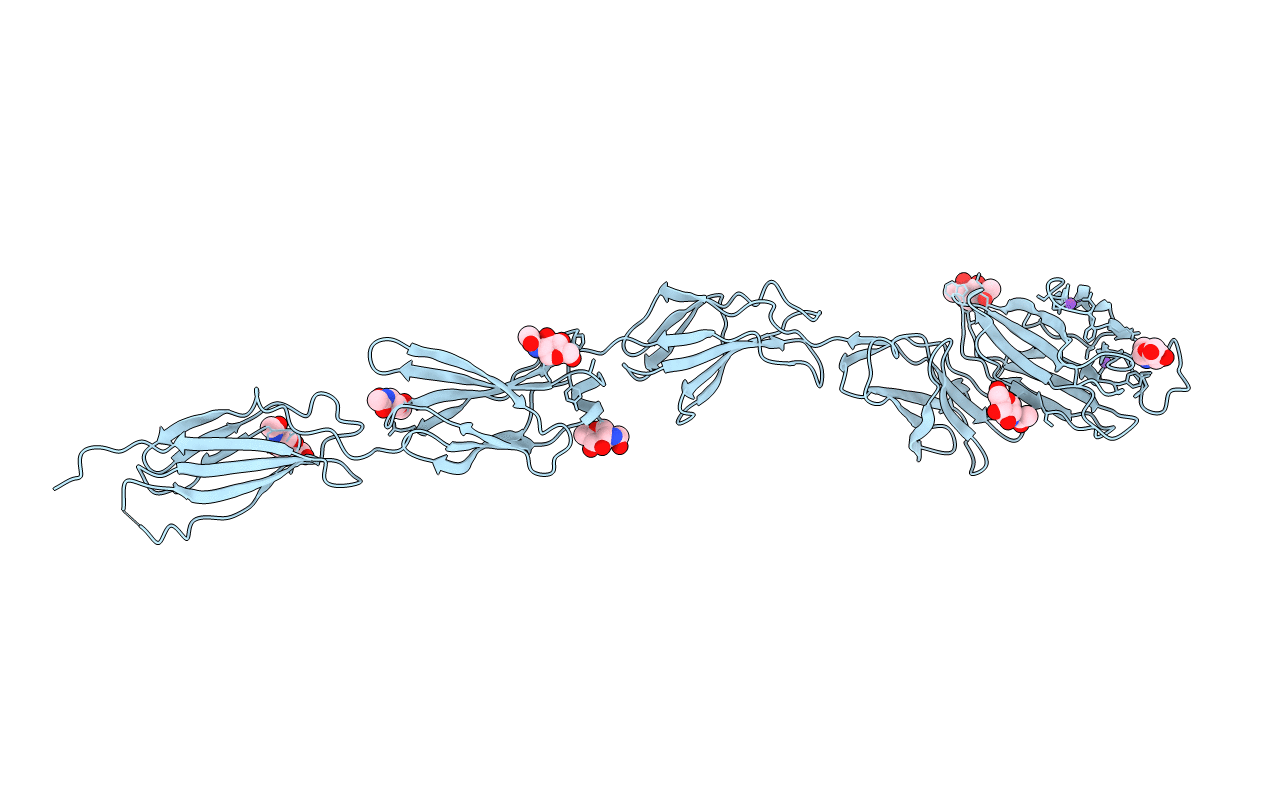
Crystal structure of the receptor protein tyrosine phosphatase mu ectodomain
Biological Source:
Source Organism:
HOMO SAPIENS (Taxon ID: 9606)
Host Organism:
Method Details:
Experimental Method:
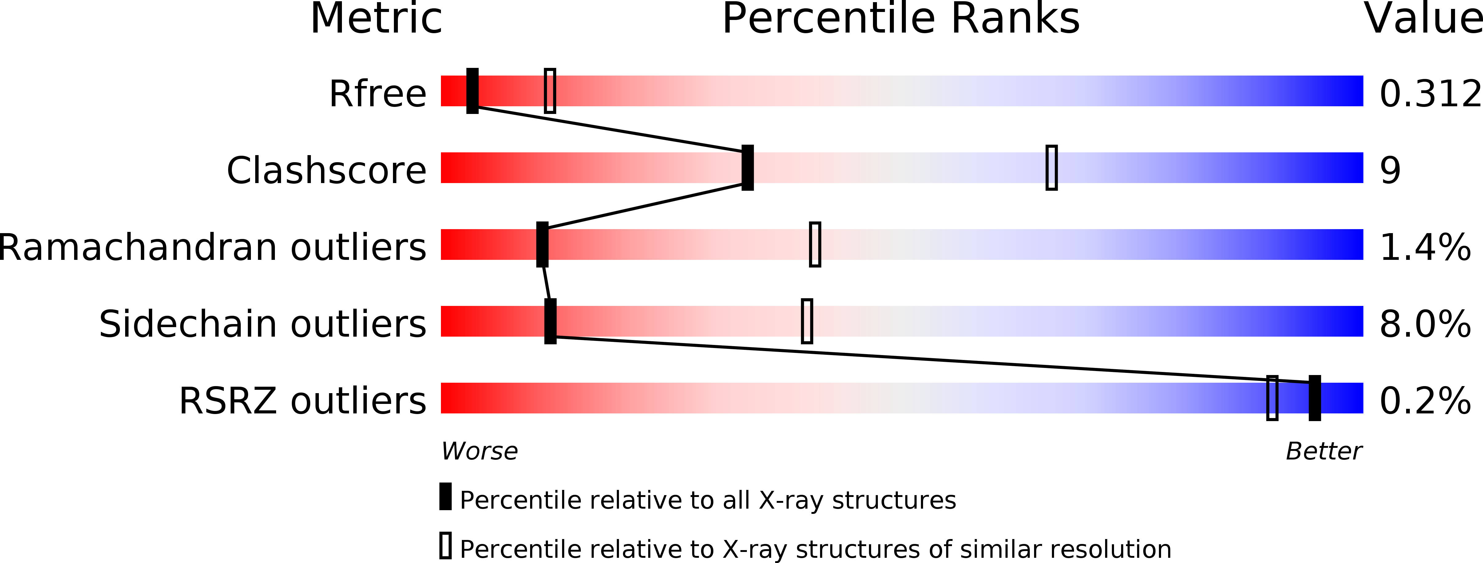
Resolution:
3.10 Å
R-Value Free:
0.32
R-Value Work:
0.24
R-Value Observed:
0.24
Space Group:
C 1 2 1
Macromolecular Entities
Structures with similar UniProt ID
Protein Blast
Polymer Type:polypeptide(L)
Molecule:RECEPTOR-TYPE TYROSINE-PROTEIN PHOSPHATASE MU
Gene (Uniprot):PTPRM
Chain IDs:A
Chain Length:731
Number of Molecules:1
Biological Source:HOMO SAPIENS
Modified Residue
Compound ID Chain ID Parent Comp ID Details 2D Image
ASN A ASN GLYCOSYLATION SITE
Primary Citation
Structure of a Tyrosine Phosphatase Adhesive Interaction Reveals a Spacer-Clamp Mechanism.
Science 317 1217 ? (2007)
PMID: 17761881 DOI: 10.1126/SCIENCE.1144646

Abstact

Cell-cell contacts are fundamental to multicellular organisms and are subject to exquisite levels of control. Human RPTPmu is a type IIB receptor protein tyrosine phosphatase that both forms an adhesive contact itself and is involved in regulating adhesion by dephosphorylating components of cadherin-catenin complexes. Here we describe a 3.1 angstrom crystal structure of the RPTPmu ectodomain that forms a homophilic trans (antiparallel) dimer with an extended and rigid architecture, matching the dimensions of adherens junctions. Cell surface expression of deletion constructs induces intercellular spacings that correlate with the ectodomain length. These data suggest that the RPTPmu ectodomain acts as a distance gauge and plays a key regulatory function, locking the phosphatase to its appropriate functional location.

Legend

Protein

Chemical

Disease

Primary Citation of related structures
Feedback Form
Name
Email
Institute
Feedback