2V5W image
Entry Detail
PDB ID:
2V5W
Title:
Crystal structure of HDAC8-substrate complex
Biological Source:
Host Organism:
PDB Version:
Deposition Date:
2007-07-10
Release Date:
2007-09-04
Method Details:
Experimental Method:
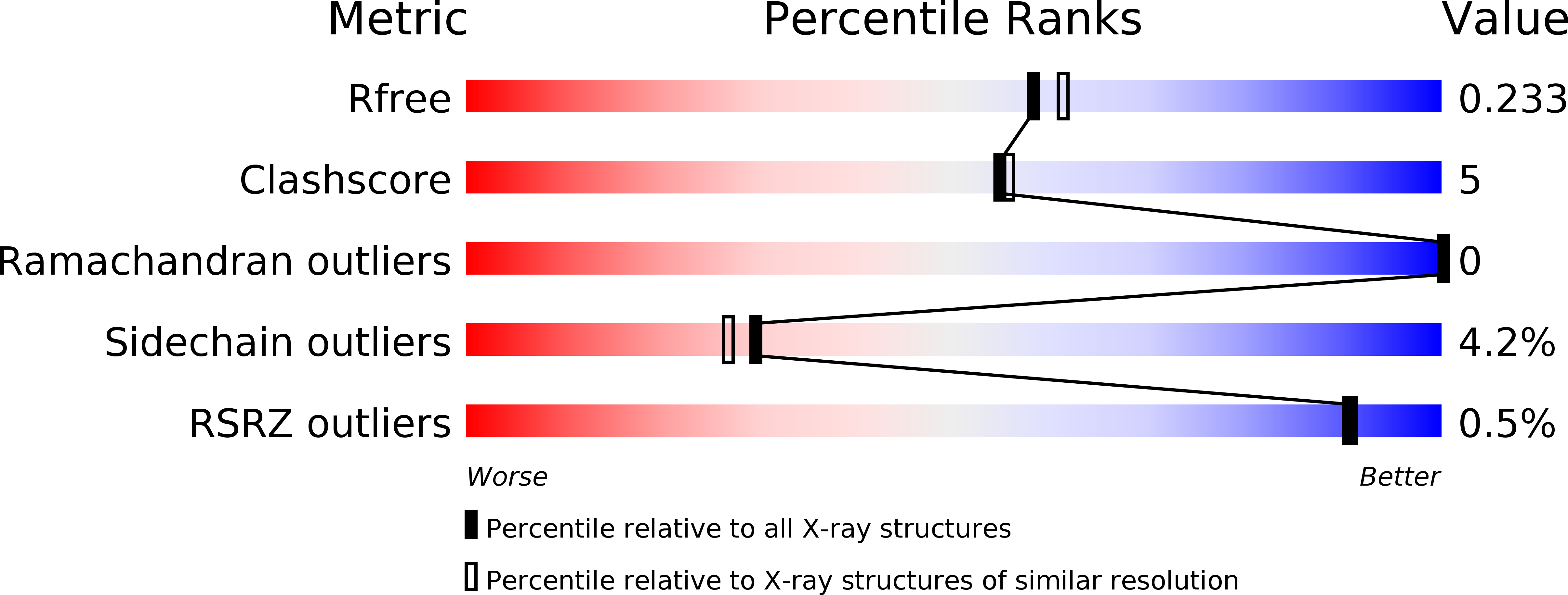
Resolution:
2.00 Å
R-Value Free:
0.23
R-Value Work:
0.17
R-Value Observed:
0.17
Space Group:
P 1 21 1
Macromolecular Entities
Polymer Type:polypeptide(L)
Description:HISTONE DEACETYLASE 8
Mutations:YES
Chain IDs:A, B
Chain Length:388
Number of Molecules:2
Biological Source:HOMO SAPIENS
Polymer Type:polypeptide(L)
Description:GLYCYL-GLYCYL-GLYCINE
Chain IDs:C (auth: G)
Chain Length:3
Number of Molecules:1
Biological Source:SYNTHETIC CONSTRUCT
Polymer Type:polypeptide(L)
Description:PEPTIDIC SUBSTRATE
Chain IDs:D (auth: I), E (auth: L)
Chain Length:6
Number of Molecules:2
Biological Source:SYNTHETIC CONSTRUCT
Modified Residue
Compound ID Chain ID Parent Comp ID Details 2D Image
ALY D LYS N(6)-ACETYLLYSINE
Primary Citation
Substrate Binding to Histone Deacetylases as Revealed by Crystal Structure of Hdac8-Substrate Complex
Embo Rep. 8 879 ? (2007)
PMID: 17721440 DOI: 10.1038/SJ.EMBOR.7401047

Abstact

Histone deacetylases (HDACs)-an enzyme family that deacetylates histones and non-histone proteins-are implicated in human diseases such as cancer, and the first-generation of HDAC inhibitors are now in clinical trials. Here, we report the 2.0 A resolution crystal structure of a catalytically inactive HDAC8 active-site mutant, Tyr306Phe, bound to an acetylated peptidic substrate. The structure clarifies the role of active-site residues in the deacetylation reaction and substrate recognition. Notably, the structure shows the unexpected role of a conserved residue at the active-site rim, Asp 101, in positioning the substrate by directly interacting with the peptidic backbone and imposing a constrained cis-conformation. A similar interaction is observed in a new hydroxamate inhibitor-HDAC8 structure that we also solved. The crucial role of Asp 101 in substrate and inhibitor recognition was confirmed by activity and binding assays of wild-type HDAC8 and Asp101Ala, Tyr306Phe and Asp101Ala/Tyr306Phe mutants.

Legend

Protein

Chemical

Disease

Primary Citation of related structures