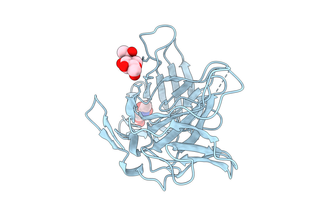
2V5N image
Entry Detail
PDB ID:
2V5N
Keywords:
Title:
STRUCTURE OF HUMAN IGF2R DOMAINS 11-12
Biological Source:
Source Organism:
Host Organism:
PDB Version:
Deposition Date:
2007-07-06
Release Date:
2007-12-11
Method Details:
Experimental Method:
Resolution:
3.20 Å
R-Value Free:
0.31
R-Value Work:
0.25
R-Value Observed:
0.25
Space Group:
P 61 2 2
Macromolecular Entities
Polymer Type:polypeptide(L)
Description:CATION-INDEPENDENT MANNOSE-6-PHOSPHATE RECEPTOR
Chain IDs:A
Chain Length:299
Number of Molecules:1
Biological Source:Homo sapiens
Modified Residue
Compound ID Chain ID Parent Comp ID Details 2D Image
ASN A ASN GLYCOSYLATION SITE
MSE A MET SELENOMETHIONINE
Ligand Molecules
Primary Citation
Structure and Functional Analysis of the Igf-II/Igf2R Interaction
Embo J. 27 265 ? (2008)
PMID: 18046459 DOI: 10.1038/SJ.EMBOJ.7601938

Abstact

Embryonic development and normal growth require exquisite control of insulin-like growth factors (IGFs). In mammals the extracellular region of the cation-independent mannose-6-phosphate receptor has gained an IGF-II-binding function and is termed type II IGF receptor (IGF2R). IGF2R sequesters IGF-II; imbalances occur in cancers and IGF2R is implicated in tumour suppression. We report crystal structures of IGF2R domains 11-12, 11-12-13-14 and domains 11-12-13/IGF-II complex. A distinctive juxtaposition of these domains provides the IGF-II-binding unit, with domain 11 directly interacting with IGF-II and domain 13 modulating binding site flexibility. Our complex shows that Phe19 and Leu53 of IGF-II lock into a hydrophobic pocket unique to domain 11 of mammalian IGF2Rs. Mutagenesis analyses confirm this IGF-II 'binding-hotspot', revealing that IGF-binding proteins and IGF2R have converged on the same high-affinity site.

Legend

Protein

Chemical

Disease

Primary Citation of related structures