2V5E image
Deposition Date 2008-10-03
Release Date 2008-10-21
Last Version Date 2024-11-13
Entry Detail
PDB ID:
2V5E
Title:
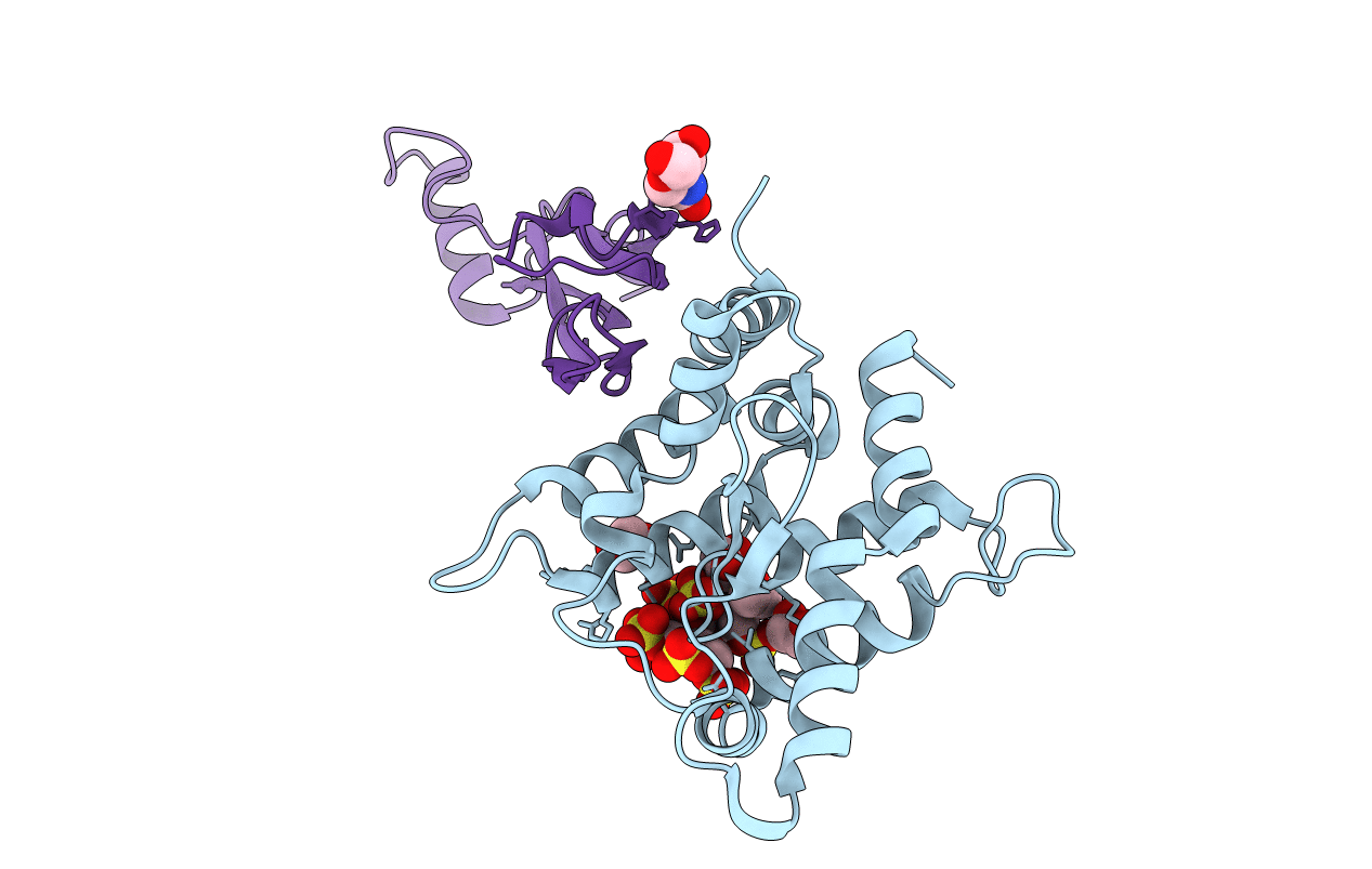
The structure of the GDNF:Coreceptor complex: Insights into RET signalling and heparin binding.
Biological Source:
Method Details:
Experimental Method:
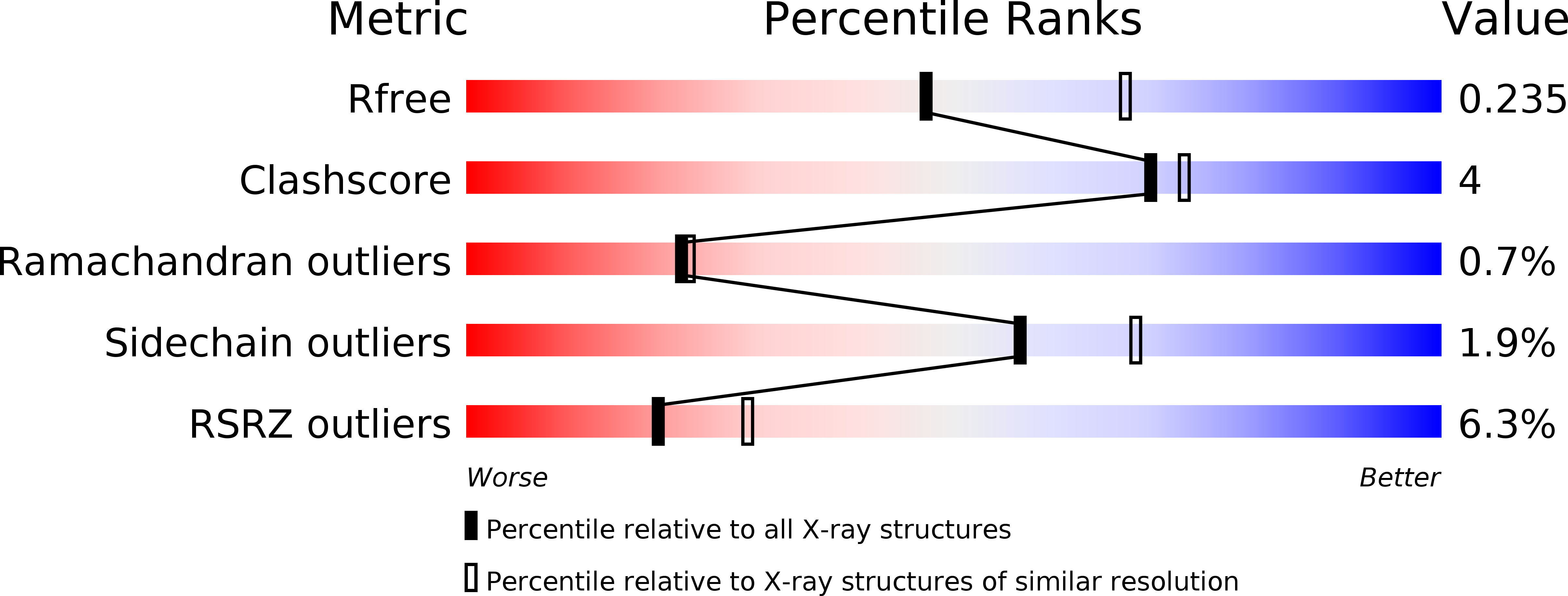
Resolution:
2.35 Å
R-Value Free:
0.23
R-Value Work:
0.18
R-Value Observed:
0.18
Space Group:
C 1 2 1
Macromolecular Entities
Polymer Type:polypeptide(L)
Molecule:GDNF FAMILY RECEPTOR ALPHA-1
Gene (Uniprot):Gfra1
Chain IDs:A
Chain Length:200
Number of Molecules:1
Biological Source:RATTUS NORVEGICUS
Polymer Type:polypeptide(L)
Molecule:GLIAL CELL LINE-DERIVED NEUROTROPHIC FACTOR
Gene (Uniprot):GDNF
Chain IDs:B
Chain Length:101
Number of Molecules:1
Biological Source:HOMO SAPIENS
Peptide-like Molecules
PRD_900013
Primary Citation
The Structure of the Glial Cell Line-Derived Neurotrophic Factor-Coreceptor Complex: Insights Into Ret Signaling and Heparin Binding.
J.Biol.Chem. 283 35164 ? (2008)
PMID: 18845535 DOI: 10.1074/JBC.M802543200

Abstact

Glial cell line-derived neurotrophic factor (GDNF), a neuronal survival factor, binds its co-receptor GDNF family receptor alpha1 (GFR alpha 1) in a 2:2 ratio and signals through the receptor tyrosine kinase RET. We have solved the GDNF(2).GFR alpha 1(2) complex structure at 2.35 A resolution in the presence of a heparin mimic, sucrose octasulfate. The structure of our GDNF(2).GFR alpha 1(2) complex and the previously published artemin(2).GFR alpha 3(2) complex are unlike in three ways. First, we have experimentally identified residues that differ in the ligand-GFR alpha interface between the two structures, in particular ones that buttress the key conserved Arg(GFR alpha)-Glu(ligand)-Arg(GFR alpha) interaction. Second, the flexible GDNF ligand "finger" loops fit differently into the GFR alphas, which are rigid. Third, and we believe most importantly, the quaternary structure of the two tetramers is dissimilar, because the angle between the two GDNF monomers is different. This suggests that the RET-RET interaction differs in different ligand(2)-co-receptor(2)-RET(2) heterohexamer complexes. Consistent with this, we showed that GDNF(2).GFR alpha1(2) and artemin(2).GFR alpha 3(2) signal differently in a mitogen-activated protein kinase assay. Furthermore, we have shown by mutagenesis and enzyme-linked immunosorbent assays of RET phosphorylation that RET probably interacts with GFR alpha 1 residues Arg-190, Lys-194, Arg-197, Gln-198, Lys-202, Arg-257, Arg-259, Glu-323, and Asp-324 upon both domains 2 and 3. Interestingly, in our structure, sucrose octasulfate also binds to the Arg(190)-Lys(202) region in GFR alpha 1 domain 2. This may explain how GDNF.GFR alpha 1 can mediate cell adhesion and how heparin might inhibit GDNF signaling through RET.

Legend

Protein

Chemical

Disease

Primary Citation of related structures