Deposition Date
2008-10-01
Release Date
2008-11-25
Last Version Date
2024-05-08
Entry Detail
PDB ID:
2V52
Keywords:
Title:
Structure of MAL-RPEL2 complexed to G-actin
Biological Source:
Source Organism(s):
MUS MUSCULUS (Taxon ID: 10090)
ORYCTOLAGUS CUNICULUS (Taxon ID: 9986)
ORYCTOLAGUS CUNICULUS (Taxon ID: 9986)
Method Details:
Experimental Method:
Resolution:
1.45 Å
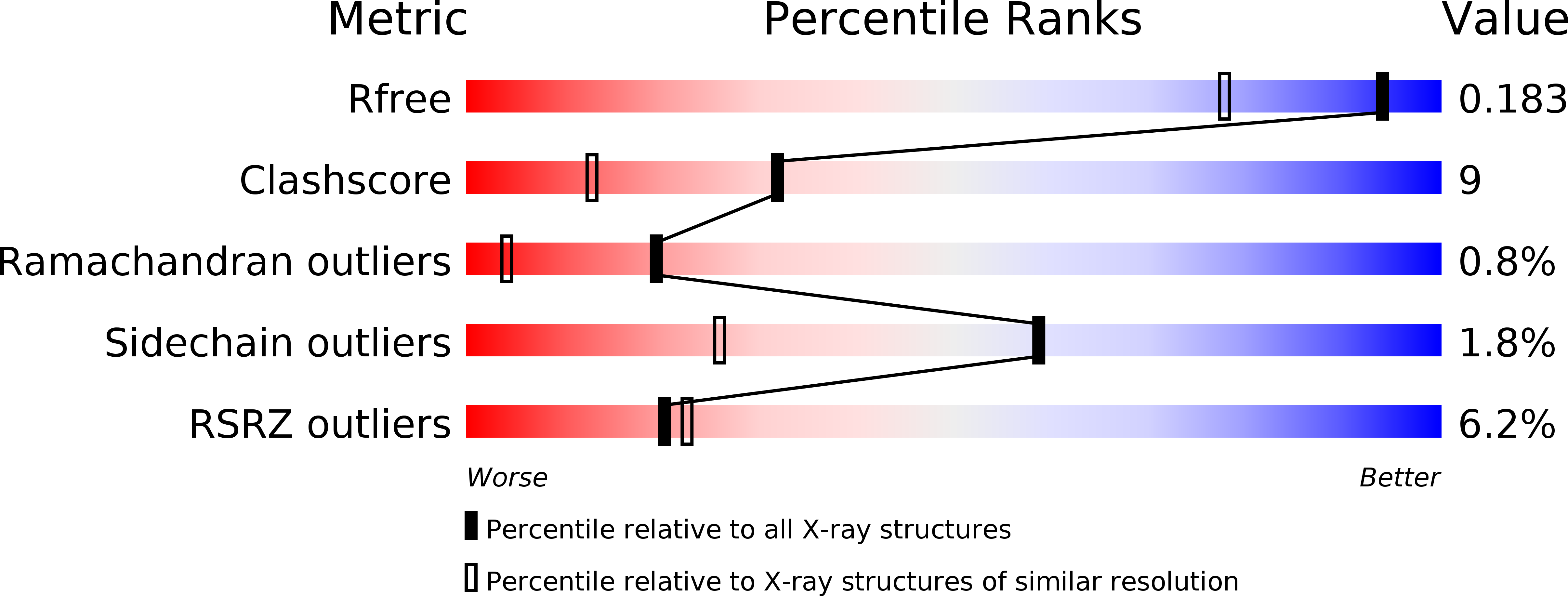
R-Value Free:
0.18
R-Value Work:
0.14
R-Value Observed:
0.14
Space Group:
P 21 21 21