2V4Z image
Deposition Date 2008-09-30
Release Date 2008-11-04
Last Version Date 2023-12-13
Entry Detail
PDB ID:
2V4Z
Keywords:
Title:
The crystal structure of the human G-protein subunit alpha (GNAI3) in complex with an engineered regulator of G-protein signaling type 2 domain (RGS2)
Biological Source:
Source Organism:
HOMO SAPIENS (Taxon ID: 9606)
Host Organism:
Method Details:
Experimental Method:
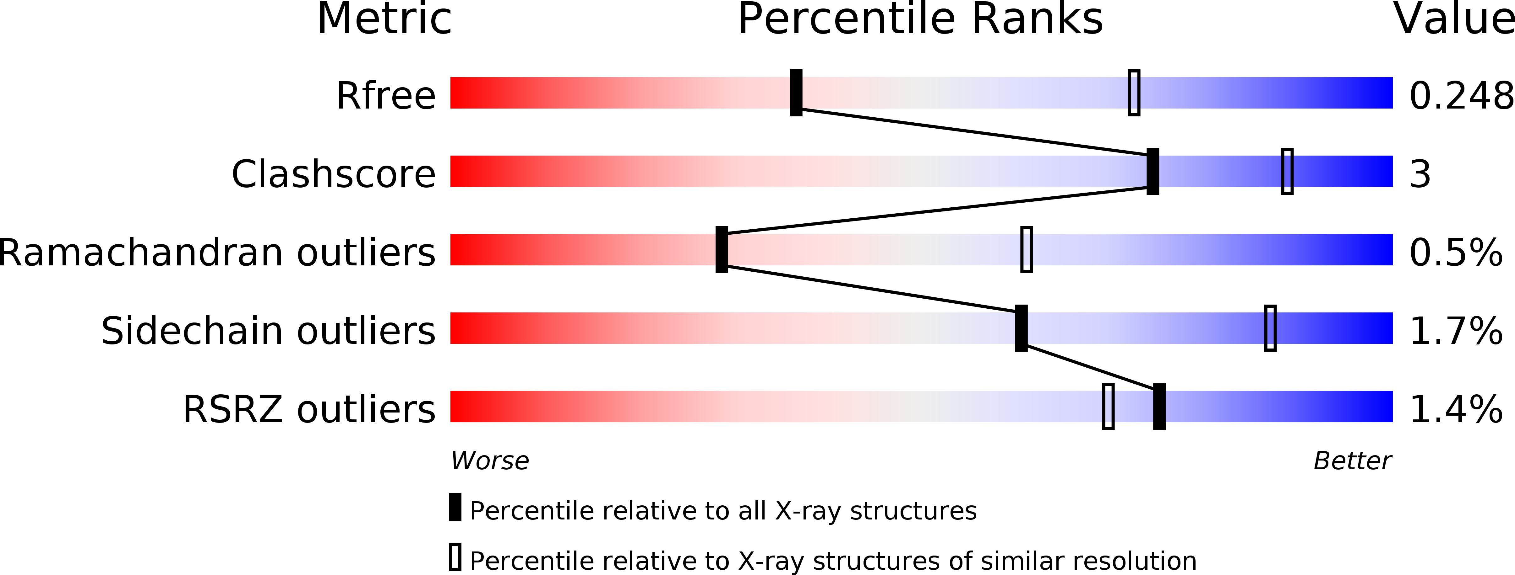
Resolution:
2.80 Å
R-Value Free:
0.25
R-Value Work:
0.20
R-Value Observed:
0.21
Space Group:
P 32 2 1
Macromolecular Entities
Structures with similar UniProt ID
Protein Blast
Polymer Type:polypeptide(L)
Molecule:GUANINE NUCLEOTIDE-BINDING PROTEIN G(K) SUBUNIT ALPHA
Gene (Uniprot):GNAI3
Chain IDs:A
Chain Length:350
Number of Molecules:1
Biological Source:HOMO SAPIENS
Structures with similar UniProt ID
Protein Blast
Polymer Type:polypeptide(L)
Molecule:REGULATOR OF G-PROTEIN SIGNALING 2
Gene (Uniprot):RGS2
Mutagens:YES
Chain IDs:B
Chain Length:142
Number of Molecules:1
Biological Source:HOMO SAPIENS
Primary Citation
Structural Determinants of G-Protein Alpha Subunit Selectivity by Regulator of G-Protein Signaling 2(Rgs2).
J.Biol.Chem. 284 19402 ? (2009)
PMID: 19478087 DOI: 10.1074/JBC.M109.024711

Abstact

"Regulator of G-protein signaling" (RGS) proteins facilitate the termination of G protein-coupled receptor (GPCR) signaling via their ability to increase the intrinsic GTP hydrolysis rate of Galpha subunits (known as GTPase-accelerating protein or "GAP" activity). RGS2 is unique in its in vitro potency and selectivity as a GAP for Galpha(q) subunits. As many vasoconstrictive hormones signal via G(q) heterotrimer-coupled receptors, it is perhaps not surprising that RGS2-deficient mice exhibit constitutive hypertension. However, to date the particular structural features within RGS2 determining its selectivity for Galpha(q) over Galpha(i/o) substrates have not been completely characterized. Here, we examine a trio of point mutations to RGS2 that elicits Galpha(i)-directed binding and GAP activities without perturbing its association with Galpha(q). Using x-ray crystallography, we determined a model of the triple mutant RGS2 in complex with a transition state mimetic form of Galpha(i) at 2.8-A resolution. Structural comparison with unliganded, wild type RGS2 and of other RGS domain/Galpha complexes highlighted the roles of these residues in wild type RGS2 that weaken Galpha(i) subunit association. Moreover, these three amino acids are seen to be evolutionarily conserved among organisms with modern cardiovascular systems, suggesting that RGS2 arose from the R4-subfamily of RGS proteins to have specialized activity as a potent and selective Galpha(q) GAP that modulates cardiovascular function.

Legend

Protein

Chemical

Disease

Primary Citation of related structures
Feedback Form
Name
Email
Institute
Feedback