2V4J image
Deposition Date 2008-09-22
Release Date 2008-12-02
Last Version Date 2024-10-23
Entry Detail
PDB ID:
2V4J
Keywords:
Title:
THE CRYSTAL STRUCTURE OF Desulfovibrio vulgaris DISSIMILATORY SULFITE REDUCTASE BOUND TO DsrC PROVIDES NOVEL INSIGHTS INTO THE MECHANISM OF SULFATE RESPIRATION
Biological Source:
Source Organism:
Method Details:
Experimental Method:
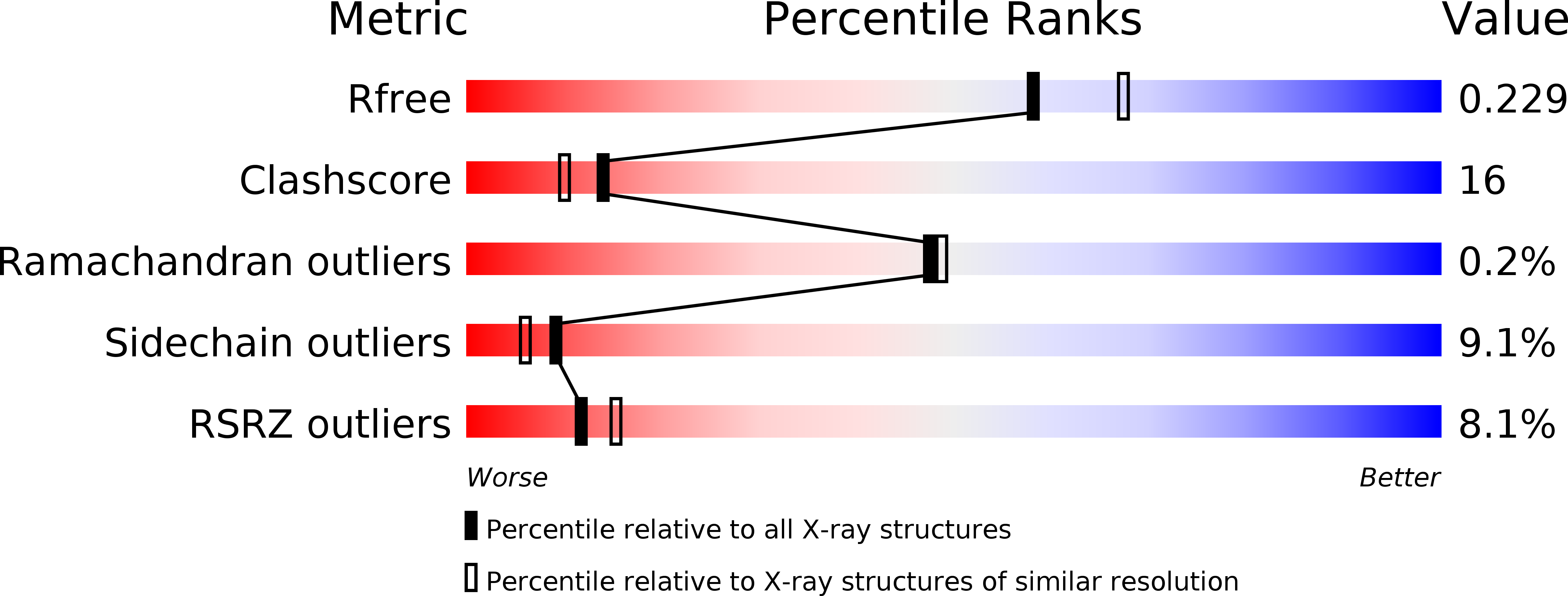
Resolution:
2.10 Å
R-Value Free:
0.21
R-Value Work:
0.19
R-Value Observed:
0.19
Space Group:
P 1 21 1
Macromolecular Entities
Structures with similar UniProt ID
Protein Blast
Polymer Type:polypeptide(L)
Molecule:SULFITE REDUCTASE, DISSIMILATORY-TYPE SUBUNIT ALPHA
Gene (Uniprot):dsvA
Chain IDs:A, D
Chain Length:437
Number of Molecules:2
Biological Source:DESULFOVIBRIO VULGARIS
Structures with similar UniProt ID
Protein Blast
Polymer Type:polypeptide(L)
Molecule:SULFITE REDUCTASE, DISSIMILATORY-TYPE SUBUNIT BETA
Gene (Uniprot):dsvB
Chain IDs:B, E
Chain Length:381
Number of Molecules:2
Biological Source:DESULFOVIBRIO VULGARIS
Structures with similar UniProt ID
Protein Blast
Polymer Type:polypeptide(L)
Molecule:SULFITE REDUCTASE, DISSIMILATORY-TYPE SUBUNIT GAMMA
Gene (Uniprot):dsvC
Chain IDs:C, F
Chain Length:105
Number of Molecules:2
Biological Source:DESULFOVIBRIO VULGARIS
Primary Citation
The Crystal Structure of Desulfovibrio Vulgaris Dissimilatory Sulfite Reductase Bound to Dsrc Provides Novel Insights Into the Mechanism of Sulfate Respiration.
J.Biol.Chem. 283 34141 ? (2008)
PMID: 18829451 DOI: 10.1074/JBC.M805643200

Abstact

Sulfate reduction is one of the earliest types of energy metabolism used by ancestral organisms to sustain life. Despite extensive studies, many questions remain about the way respiratory sulfate reduction is associated with energy conservation. A crucial enzyme in this process is the dissimilatory sulfite reductase (dSiR), which contains a unique siroheme-[4Fe4S] coupled cofactor. Here, we report the structure of desulfoviridin from Desulfovibrio vulgaris, in which the dSiR DsrAB (sulfite reductase) subunits are bound to the DsrC protein. The alpha(2)beta(2)gamma(2) assembly contains two siroheme-[4Fe4S] cofactors bound by DsrB, two sirohydrochlorins and two [4Fe4S] centers bound by DsrA, and another four [4Fe4S] centers in the ferredoxin domains. A sulfite molecule, coordinating the siroheme, is found at the active site. The DsrC protein is bound in a cleft between DsrA and DsrB with its conserved C-terminal cysteine reaching the distal side of the siroheme. We propose a novel mechanism for the process of sulfite reduction involving DsrAB, DsrC, and the DsrMKJOP membrane complex (a membrane complex with putative disulfide/thiol reductase activity), in which two of the six electrons for reduction of sulfite derive from the membrane quinone pool. These results show that DsrC is involved in sulfite reduction, which changes the mechanism of sulfate respiration. This has important implications for models used to date ancient sulfur metabolism based on sulfur isotope fractionations.

Legend

Protein

Chemical

Disease

Primary Citation of related structures
Feedback Form
Name
Email
Institute
Feedback