2V3L image
Deposition Date 2007-06-18
Release Date 2007-10-09
Last Version Date 2024-05-15
Entry Detail
PDB ID:
2V3L
Keywords:
Title:
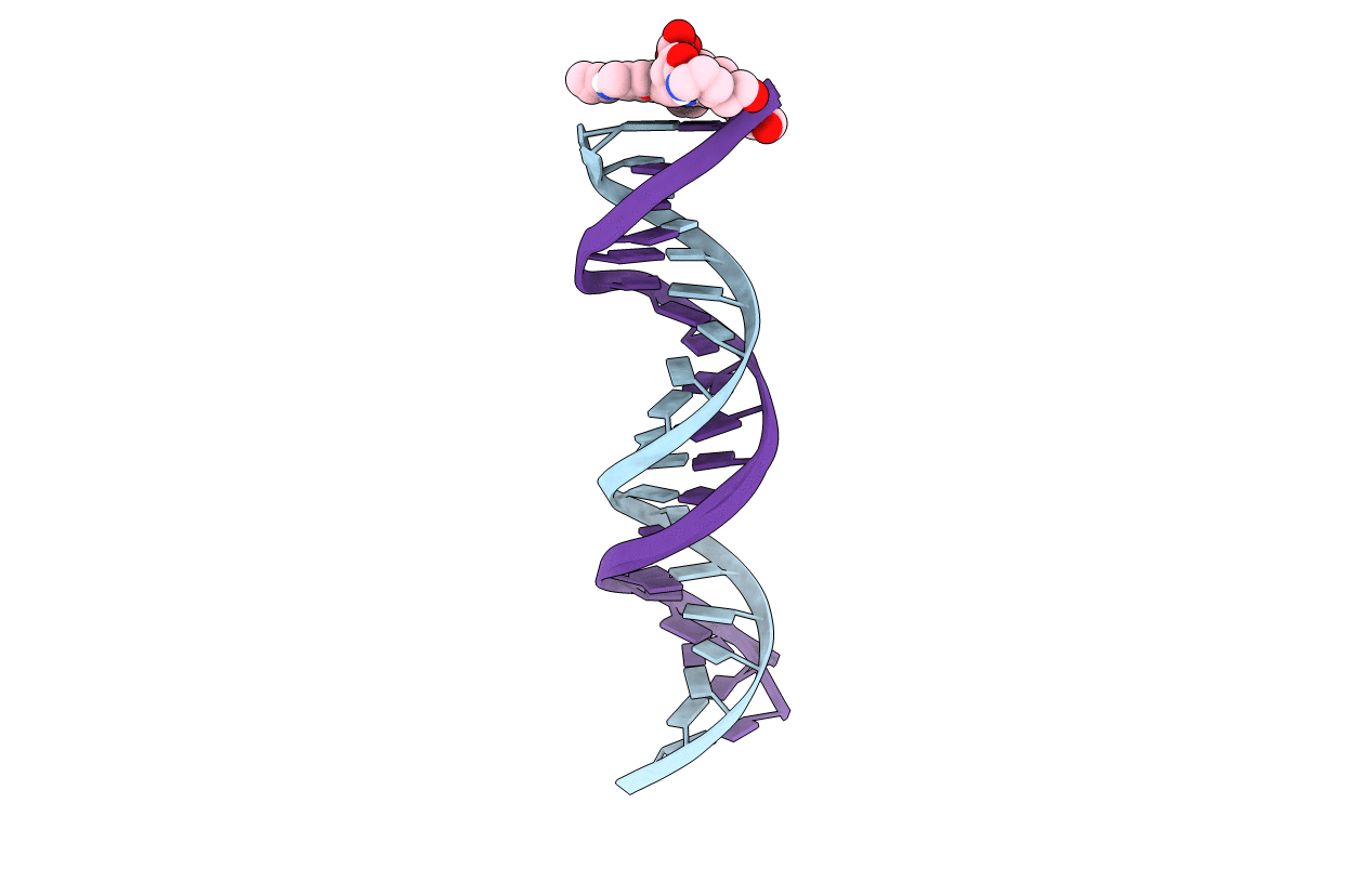
Orientational and dynamical heterogeneity of Rhodamine 6G terminally attached to a DNA helix
Biological Source:
Source Organism:
Method Details:
Experimental Method:
Conformers Submitted:
2
Macromolecular Entities
Polymer Type:polydeoxyribonucleotide
Molecule:5'-D(*GP*AP*AP*TP*GP*GP*CP*GP*AP*AP *TP*GP*GP*CP*GP*CP*TP*TP*TP*G)-3'
Chain IDs:A
Chain Length:20
Number of Molecules:1
Biological Source:synthetic construct
Polymer Type:polydeoxyribonucleotide
Molecule:5'-D(*CP*AP*AP*AP*GP*CP*GP*CP*CP*AP *TP*TP*CP*GP*CP*CP*AP*TP*TP*C)-3'
Chain IDs:B
Chain Length:20
Number of Molecules:1
Biological Source:synthetic construct
Ligand Molecules
Primary Citation
Orientational and Dynamical Heterogeneity of Rhodamine 6G Terminally Attached to a DNA Helix Revealed by NMR and Single-Molecule Fluorescence Spectroscopy.
J.Am.Chem.Soc. 129 12746 ? (2007)
PMID: 17900110 DOI: 10.1021/JA0722574

Abstact

The comparison of Förster resonance energy transfer (FRET) efficiencies between two fluorophores covalently attached to a single protein or DNA molecule is an elegant approach for deducing information about their structural and dynamical heterogeneity. For a more detailed structural interpretation of single-molecule FRET assays, information about the positions as well as the dynamics of the dye labels attached to the biomolecule is important. In this work, Rhodamine 6G (2-[3'-(ethylamino)-6'-(ethylimino)-2',7'-dimethyl-6'H-xanthen-9'-yl]-benzoic acid) bound to the 5'-end of a 20 base pair long DNA duplex is investigated by both single-molecule multiparameter fluorescence detection (MFD) experiments and NMR spectroscopy. Rhodamine 6G is commonly employed in nucleic acid research as a FRET dye. MFD experiments directly reveal the equilibrium of the dye bound to DNA between three heterogeneous environments, which are characterized by distinct fluorescence lifetime and intensity distributions as a result of different guanine-dye excited-state electron transfer interactions. Sub-ensemble fluorescence autocorrelation analysis shows the highly dynamic character of the dye-DNA interactions ranging from nano- to milliseconds and species-specific triplet relaxation times. Two-dimensional NMR spectroscopy corroborates this information by the determination of the detailed geometric structures of the dye-nucleobase complex and their assignment to each population observed in the single-molecule fluorescence experiments. From both methods, a consistent and detailed molecular description of the structural and dynamical heterogeneity is obtained.

Legend

Protein

Chemical

Disease

Primary Citation of related structures