2V2G image
Deposition Date 2007-06-05
Release Date 2008-04-08
Last Version Date 2023-12-13
Entry Detail
PDB ID:
2V2G
Keywords:
Title:
Crystal Structure of the C45S mutant of the Peroxiredoxin 6 of Arenicola Marina. Monoclinic form
Biological Source:
Source Organism:
Host Organism:
Method Details:
Experimental Method:
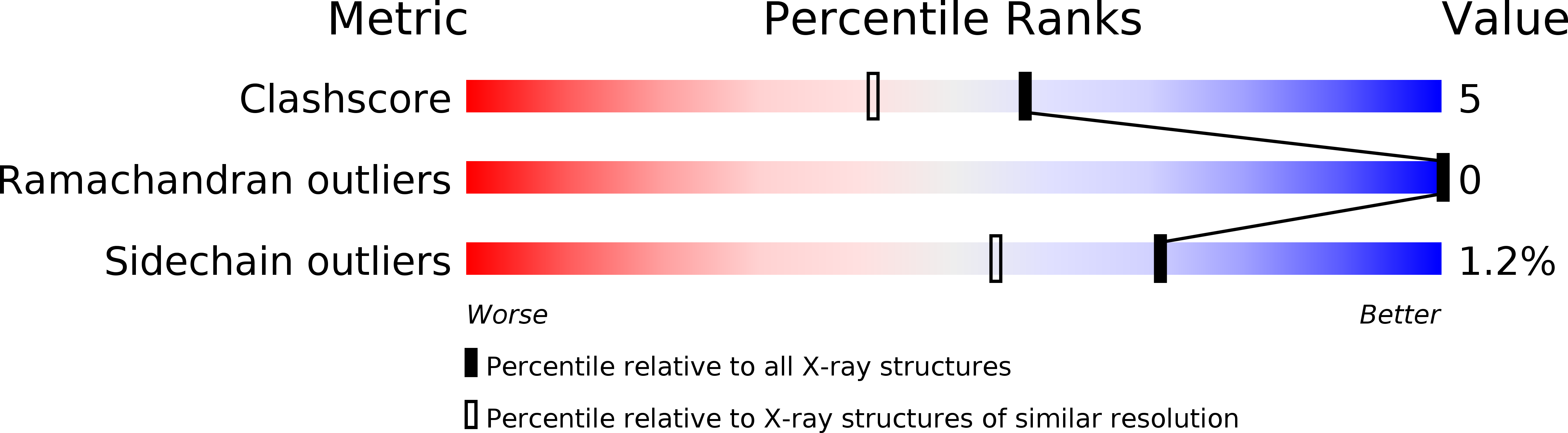
Resolution:
1.60 Å
R-Value Free:
0.19
R-Value Work:
0.16
R-Value Observed:
0.16
Space Group:
C 1 2 1
Macromolecular Entities
Polymer Type:polypeptide(L)
Molecule:PEROXIREDOXIN 6
Gene (Uniprot):Prdx6
Mutations:YES
Chain IDs:A, B, C, D
Chain Length:233
Number of Molecules:4
Biological Source:ARENICOLA MARINA
Primary Citation
The Crystal Structure of the C45S Mutant of Annelid Arenicola Marina Peroxiredoxin 6 Supports its Assignment to the Mechanistically Typical 2- Cys Subfamily without Any Formation of Toroid- Shaped Decamers.
Protein Sci. 17 700 ? (2008)
PMID: 18359859 DOI: 10.1110/PS.073399308

Abstact

The peroxiredoxins (PRDXs) define a superfamily of thiol-dependent peroxidases able to reduce hydrogen peroxide, alkyl hydroperoxides, and peroxynitrite. Besides their cytoprotective antioxidant function, PRDXs have been implicated in redox signaling and chaperone activity, the latter depending on the formation of decameric high-molecular-weight structures. PRDXs have been mechanistically divided into three major subfamilies, namely typical 2-Cys, atypical 2-Cys, and 1-Cys PRDXs, based on the number and position of cysteines involved in the catalysis. We report the structure of the C45S mutant of annelid worm Arenicola marina PRDX6 in three different crystal forms determined at 1.6, 2.0, and 2.4 A resolution. Although A. marina PRDX6 was cloned during the search of annelid homologs of mammalian 1-Cys PRDX6s, the crystal structures support its assignment to the mechanistically typical 2-Cys PRDX subfamily. The protein is composed of two distinct domains: a C-terminal domain and an N-terminal domain exhibiting a thioredoxin fold. The subunits are associated in dimers compatible with the formation of intersubunit disulfide bonds between the peroxidatic and the resolving cysteine residues in the wild-type enzyme. The packing of two crystal forms is very similar, with pairs of dimers associated as tetramers. The toroid-shaped decamers formed by dimer association and observed in most typical 2-Cys PRDXs is not present. Thus, A. marina PRDX6 presents structural features of typical 2-Cys PRDXs without any formation of toroid-shaped decamers, suggesting that it should function more like a cytoprotective antioxidant enzyme or a modulator of peroxide-dependent cell signaling rather than a molecular chaperone.

Legend

Protein

Chemical

Disease

Primary Citation of related structures