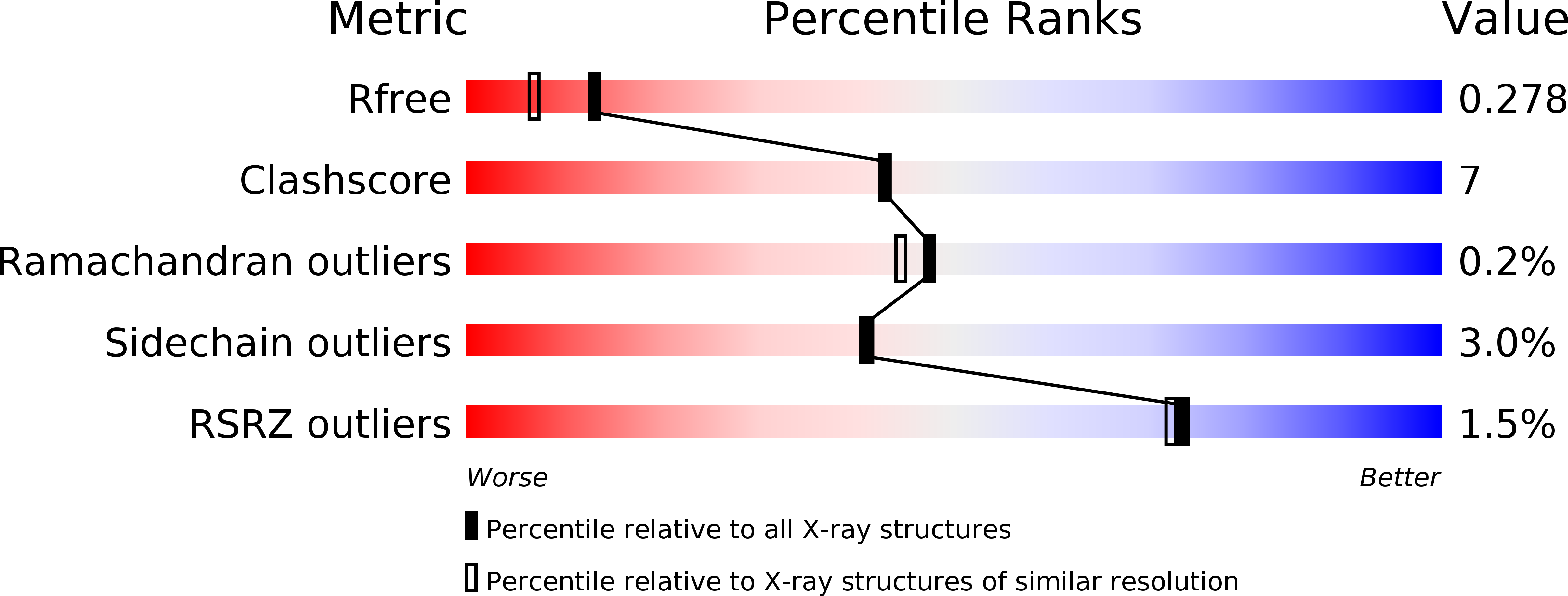
2V1X image
Deposition Date 2007-05-30
Release Date 2007-07-03
Last Version Date 2023-12-13
Entry Detail
PDB ID:
2V1X
Keywords:
Title:
Crystal structure of human RECQ-like DNA helicase
Biological Source:
Source Organism:
HOMO SAPIENS (Taxon ID: 9606)
Host Organism:
Method Details:
Experimental Method:
Resolution:
2.00 Å
R-Value Free:
0.27
R-Value Work:
0.23
R-Value Observed:
0.23
Space Group:
P 1 21 1
Macromolecular Entities
Structures with similar UniProt ID
Protein Blast
Polymer Type:polypeptide(L)
Molecule:ATP-DEPENDENT DNA HELICASE Q1
Gene (Uniprot):RECQL
Chain IDs:A, B
Chain Length:591
Number of Molecules:2
Biological Source:HOMO SAPIENS
Primary Citation
Structure of the Human Recq1 Helicase Reveals a Putative Strand-Separation Pin.
Proc.Natl.Acad.Sci.USA 106 1039 ? (2009)
PMID: 19151156 DOI: 10.1073/PNAS.0806908106

Abstact

RecQ-like helicases, which include 5 members in the human genome, are important in maintaining genome integrity. We present a crystal structure of a truncated form of the human RECQ1 protein with Mg-ADP. The truncated protein is active in DNA fork unwinding but lacks other activities of the full-length enzyme: disruption of Holliday junctions and DNA strand annealing. The structure of human RECQ1 resembles that of Escherichia coli RecQ, with some important differences. All structural domains are conserved, including the 2 RecA-like domains and the RecQ-specific zinc-binding and winged-helix (WH) domains. However, the WH domain is positioned at a different orientation from that of the E. coli enzyme. We identify a prominent beta-hairpin of the WH domain as essential for DNA strand separation, which may be analogous to DNA strand-separation features of other DNA helicases. This hairpin is significantly shorter in the E. coli enzyme and is not required for its helicase activity, suggesting that there are significant differences between the modes of action of RecQ family members.

Legend

Protein

Chemical

Disease

Primary Citation of related structures