2V1B image
Deposition Date 2007-05-22
Release Date 2007-12-11
Last Version Date 2024-11-06
Entry Detail
PDB ID:
2V1B
Keywords:
Title:
N- and C-terminal helices of oat LOV2 (404-546) are involved in light-induced signal transduction (room temperature (293K) light structure of LOV2 (404-546))
Biological Source:
Source Organism:
AVENA SATIVA (Taxon ID: 4498)
Host Organism:
Method Details:
Experimental Method:
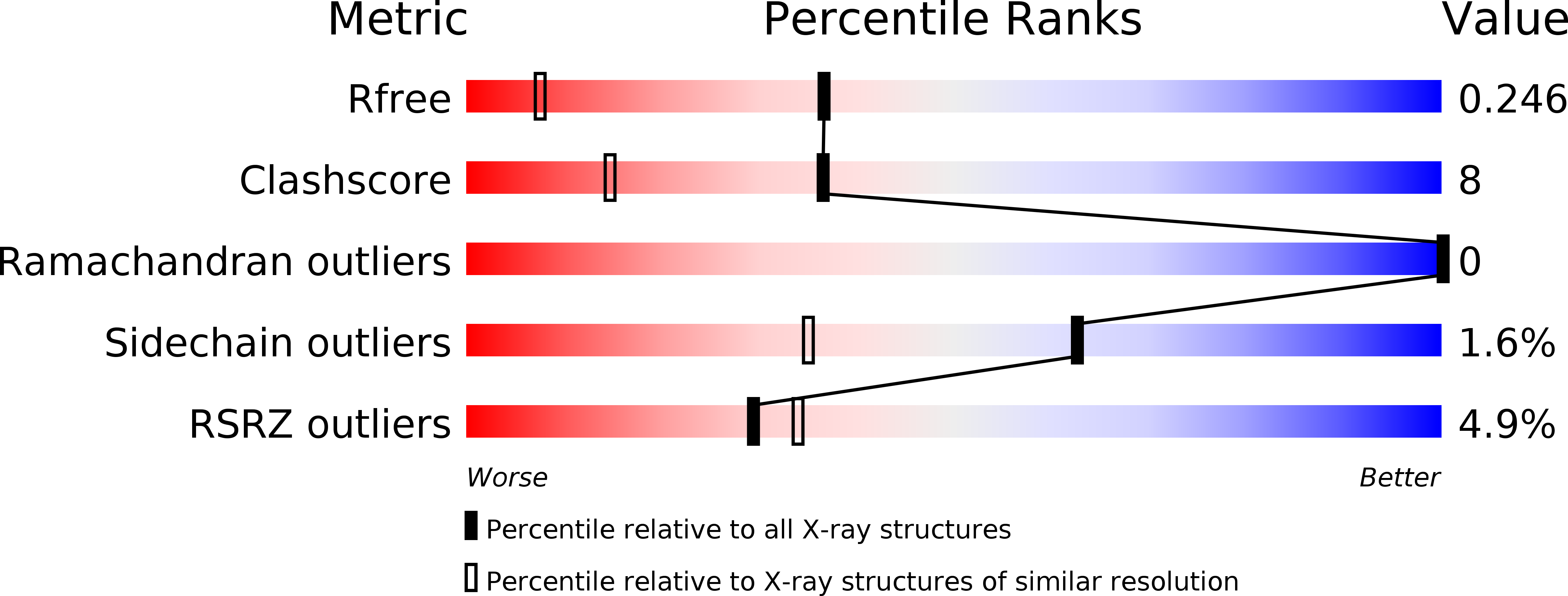
Resolution:
1.55 Å
R-Value Free:
0.21
R-Value Work:
0.16
R-Value Observed:
0.16
Space Group:
P 21 21 21
Macromolecular Entities
Polymer Type:polypeptide(L)
Molecule:NPH1-1
Gene (Uniprot):NPH1-1
Chain IDs:A
Chain Length:144
Number of Molecules:1
Biological Source:AVENA SATIVA
Primary Citation
N- and C-Terminal Flanking Regions Modulate Light-Induced Signal Transduction in the Lov2 Domain of the Blue Light Sensor Phototropin 1 from Avena Sativa.
Biochemistry 46 14001 ? (2007)
PMID: 18001137 DOI: 10.1021/BI701543E

Abstact

Light sensing by photoreceptors controls phototropism, chloroplast movement, stomatal opening, and leaf expansion in plants. Understanding the molecular mechanism by which these processes are regulated requires a quantitative description of photoreceptor dynamics. We focus on a light-driven signal transduction mechanism in the LOV2 domain (LOV, light, oxygen, voltage) of the blue light photoreceptor phototropin 1 from Avena sativa (oat). High-resolution crystal structures of the dark and light states of an oat LOV2 construct including residues Leu404 through Leu546 (LOV2 (404-546)) have been determined at 105 and 293 K. In all four structures, LOV2 (404-546) exhibits the typical Per-ARNT-Sim (PAS) fold, flanked by an additional conserved N-terminal turn-helix-turn motif and a C-terminal flanking region containing an amphipathic Jalpha helix. These regions dock on the LOV2 core domain and bury several hydrophobic residues of the central beta-sheet of the core domain that would otherwise be exposed to solvent. Light structures of LOV2 (404-546) reveal that formation of the covalent bond between Cys450 and the C4a atom of the flavin mononucleotide (FMN) results in local rearrangement of the hydrogen-bonding network in the FMN binding pocket. These rearrangements are associated with disruption of the Asn414-Asp515 hydrogen bond on the surface of the protein and displacement of the N- and C-terminal flanking regions of LOV2 (404-546), both of which constitute a structural signal.

Legend

Protein

Chemical

Disease

Primary Citation of related structures