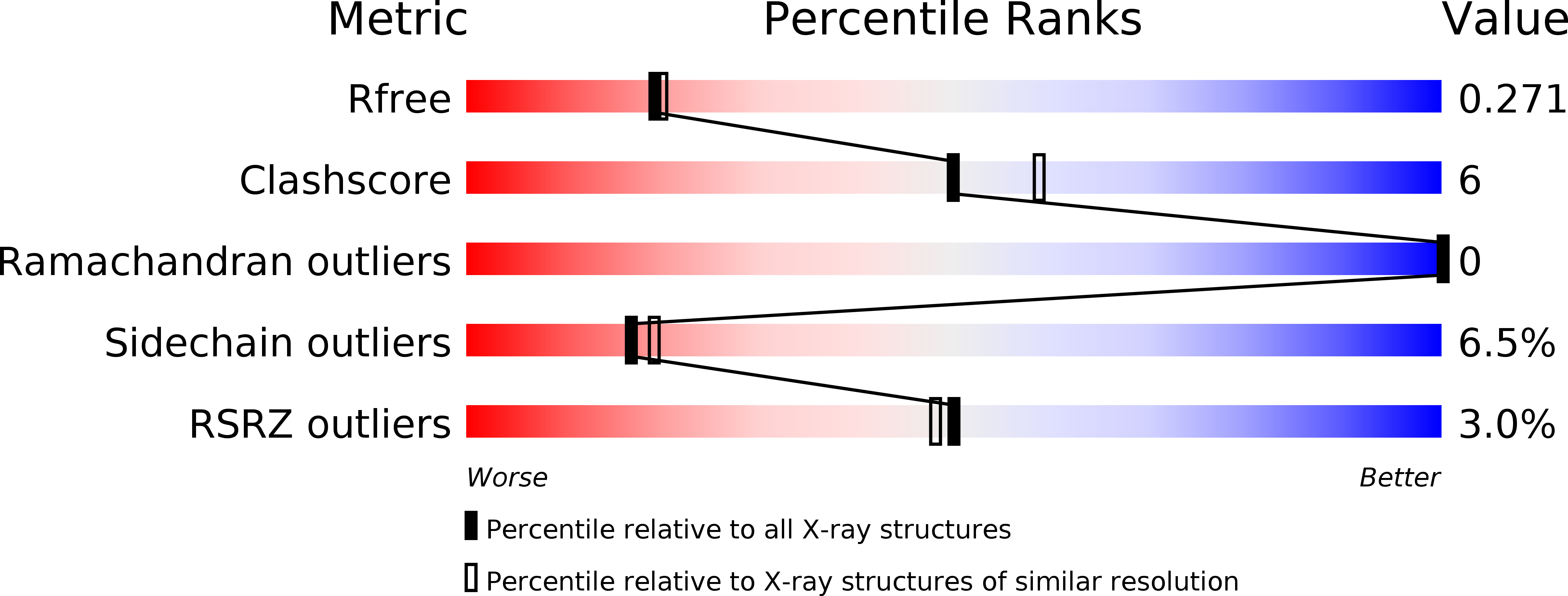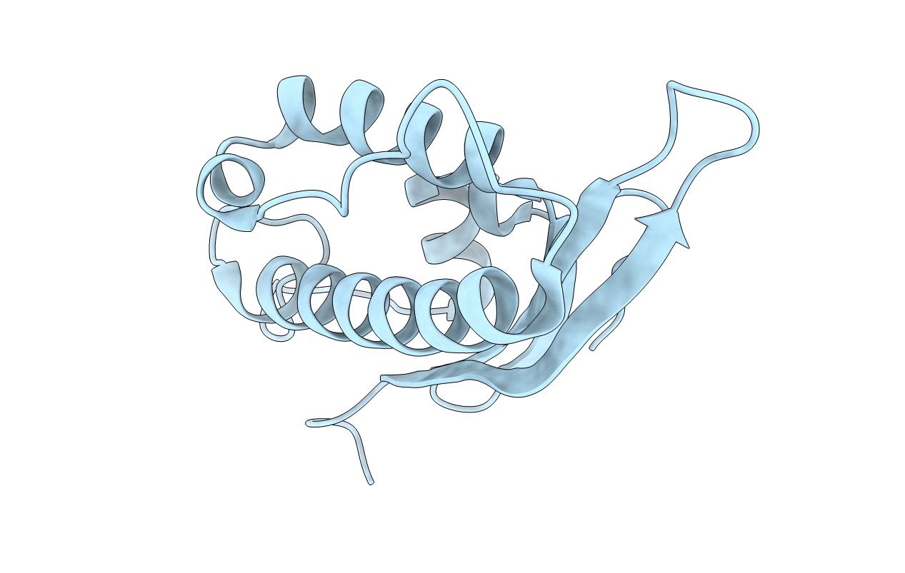
2V14 image
Deposition Date 2007-05-21
Release Date 2007-07-31
Last Version Date 2024-10-23
Entry Detail
PDB ID:
2V14
Title:
Kinesin 16B Phox-homology domain (KIF16B)
Biological Source:
Source Organism:
HOMO SAPIENS (Taxon ID: 9606)
Host Organism:
Method Details:
Experimental Method:
Resolution:
2.20 Å
R-Value Free:
0.28
R-Value Work:
0.20
R-Value Observed:
0.21
Space Group:
I 2 3
Macromolecular Entities
Structures with similar UniProt ID
Protein Blast
Polymer Type:polypeptide(L)
Molecule:KINESIN-LIKE MOTOR PROTEIN C20ORF23
Gene (Uniprot):KIF16B
Chain IDs:A
Chain Length:134
Number of Molecules:1
Biological Source:HOMO SAPIENS
Modified Residue
Compound ID Chain ID Parent Comp ID Details 2D Image
MSE A MET SELENOMETHIONINE
Primary Citation
The Structural Basis of Novel Endosome Anchoring Activity of Kif16B Kinesin.
Embo J. 26 3709 ? (2007)
PMID: 17641687 DOI: 10.1038/SJ.EMBOJ.7601800

Abstact

KIF16B is a newly identified kinesin that regulates the intracellular motility of early endosomes. KIF16B is unique among kinesins in that its cargo binding is mediated primarily by the strong interaction of its PX domain with endosomal lipids. To elucidate the structural basis of this unique endosomal anchoring activity of KIF16B-PX, we determined the crystal structure of the PX domain and performed in vitro and cellular membrane binding measurements for KIF16B-PX and mutants. The most salient structural feature of KIF16B-PX is that two neighboring residues, L1248 and F1249, on the membrane-binding surface form a protruding hydrophobic stalk with a large solvent-accessible surface area. This unique structure, arising from the complementary stacking of the two side chains and the local conformation, allows strong hydrophobic membrane interactions and endosome tethering. The presence of similar hydrophobic pairs in the amino-acid sequences of other membrane-binding domains and proteins suggests that the same structural motif may be shared by other membrane-binding proteins, whose physiological functions depend on strong hydrophobic membrane interactions.

Legend

Protein

Chemical

Disease

Primary Citation of related structures