Deposition Date
2007-05-08
Release Date
2008-06-10
Last Version Date
2024-05-08
Entry Detail
PDB ID:
2V04
Keywords:
Title:
CRYSTAL STRUCTURE OF CHOLINE BINDING PROTEIN F FROM STREPTOCOCCUS PNEUMONIAE
Biological Source:
Source Organism(s):
STREPTOCOCCUS PNEUMONIAE (Taxon ID: 171101)
Expression System(s):
Method Details:
Experimental Method:
Resolution:
2.10 Å
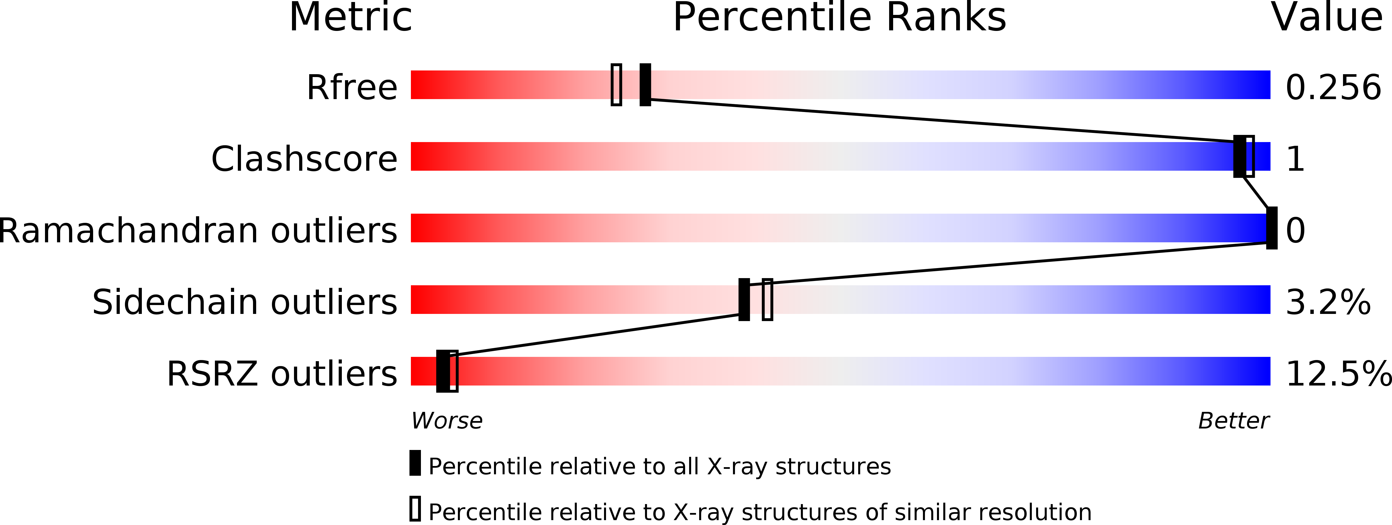
R-Value Free:
0.25
R-Value Work:
0.20
R-Value Observed:
0.21
Space Group:
P 21 21 2