2UXK image
Entry Detail
PDB ID:
2UXK
Keywords:
Title:
X-ray high resolution structure of the photosynthetic reaction center from Rb. sphaeroides at pH 10 in the charge-separated state
Biological Source:
Source Organism:
PDB Version:
Deposition Date:
2007-03-28
Release Date:
2007-07-03
Method Details:
Experimental Method:
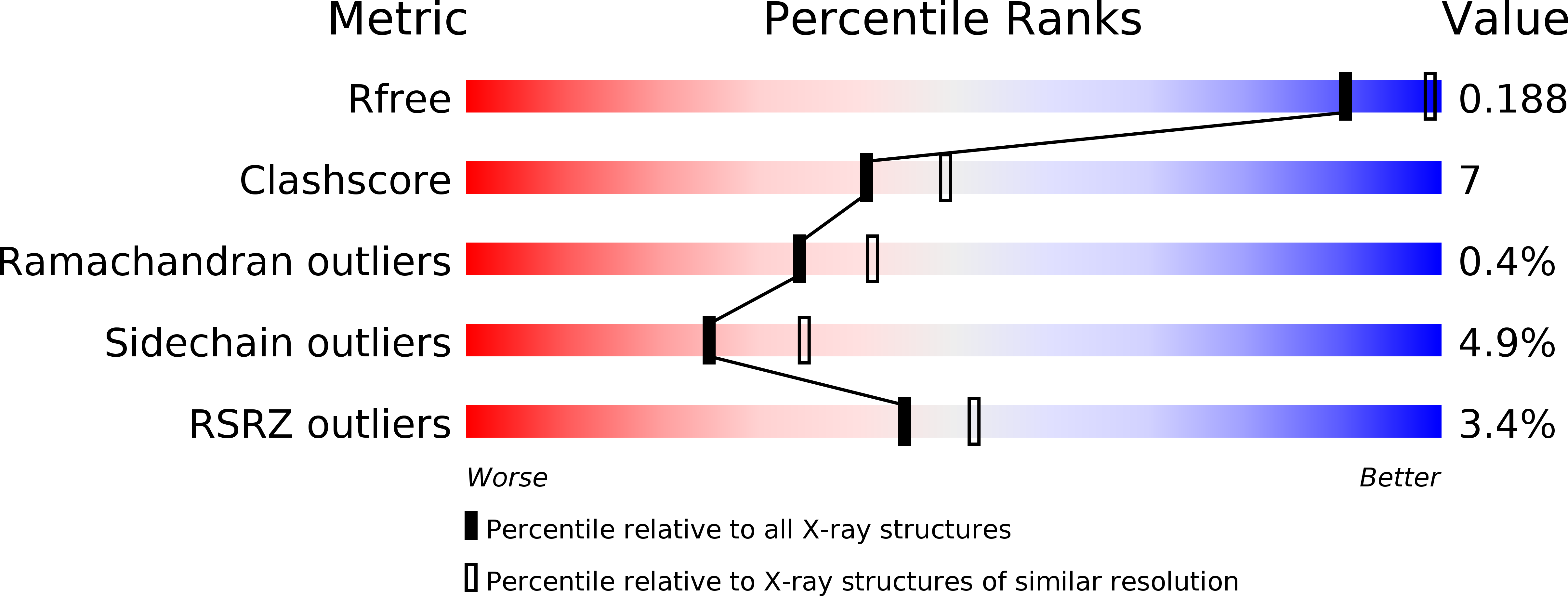
Resolution:
2.31 Å
R-Value Free:
0.21
R-Value Work:
0.19
R-Value Observed:
0.19
Space Group:
P 43 21 2
Macromolecular Entities
Polymer Type:polypeptide(L)
Description:REACTION CENTER PROTEIN H CHAIN
Chain IDs:A (auth: H)
Chain Length:260
Number of Molecules:1
Biological Source:RHODOBACTER SPHAEROIDES
Polymer Type:polypeptide(L)
Description:REACTION CENTER PROTEIN L CHAIN
Chain IDs:B (auth: L)
Chain Length:281
Number of Molecules:1
Biological Source:RHODOBACTER SPHAEROIDES
Polymer Type:polypeptide(L)
Description:REACTION CENTER PROTEIN M CHAIN
Chain IDs:C (auth: M)
Chain Length:307
Number of Molecules:1
Biological Source:RHODOBACTER SPHAEROIDES
Primary Citation
Ph Modulates the Quinone Position in the Photosynthetic Reaction Center from Rhodobacter Sphaeroides in the Neutral and Charge Separated States.
J.Mol.Biol. 371 396 ? (2007)
PMID: 17570397 DOI: 10.1016/J.JMB.2007.04.082

Abstact

The structure of the photosynthetic reaction-center from Rhodobacter sphaeroides has been determined at four different pH values (6.5, 8.0, 9.0, 10.0) in the neutral and in charge separated states. At pH 8.0, in the neutral state, we obtain a resolution of 1.87 A, which is the best ever reported for the bacterial reaction center protein. Our crystallographic data confirm the existence of two different binding positions of the secondary quinone (QB). We observe a new orientation of QB in its distal position, which shows no ring-flip compared to the orientation in the proximal position. Datasets collected for the different pH values show a pH-dependence of the population of the proximal position. The new orientation of QB in the distal position and the pH-dependence could be confirmed by continuum electrostatics calculations. Our calculations are in agreement with the experimentally observed proton uptake upon charge separation. The high resolution of our crystallographic data allows us to identify new water molecules and external residues being involved in two previously described hydrogen bond proton channels. These extended proton-transfer pathways, ending at either of the two oxo-groups of QB in its proximal position, provide additional evidence that ring-flipping is not required for complete protonation of QB upon reduction.

Legend

Protein

Chemical

Disease