
Deposition Date
2007-03-22
Release Date
2007-10-02
Last Version Date
2025-10-01
Entry Detail
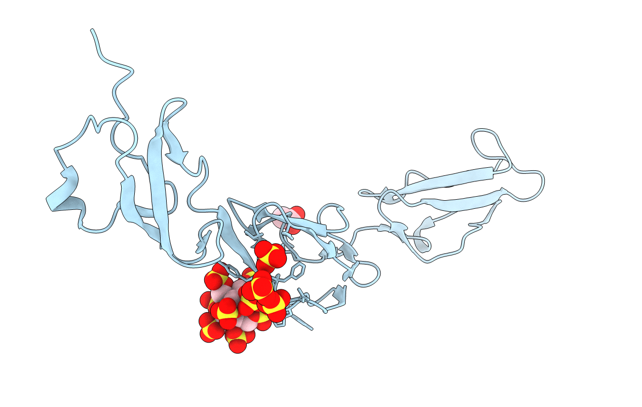
PDB ID:
2UWN
Keywords:
Title:
Crystal structure of Human Complement Factor H, SCR domains 6-8 (H402 risk variant), in complex with ligand.
Biological Source:
Source Organism(s):
HOMO SAPIENS (Taxon ID: 9606)
Expression System(s):
Method Details:
Experimental Method:
Resolution:
2.35 Å
R-Value Free:
0.24
R-Value Work:
0.21
R-Value Observed:
0.21
Space Group:
C 2 2 21
