Deposition Date
2007-03-13
Release Date
2007-08-21
Last Version Date
2024-05-08
Entry Detail
PDB ID:
2UVP
Keywords:
Title:
Crystal structure of HobA (HP1230)from Helicobacter pylori
Biological Source:
Source Organism(s):
HELICOBACTER PYLORI (Taxon ID: 210)
Expression System(s):
Method Details:
Experimental Method:
Resolution:
1.70 Å
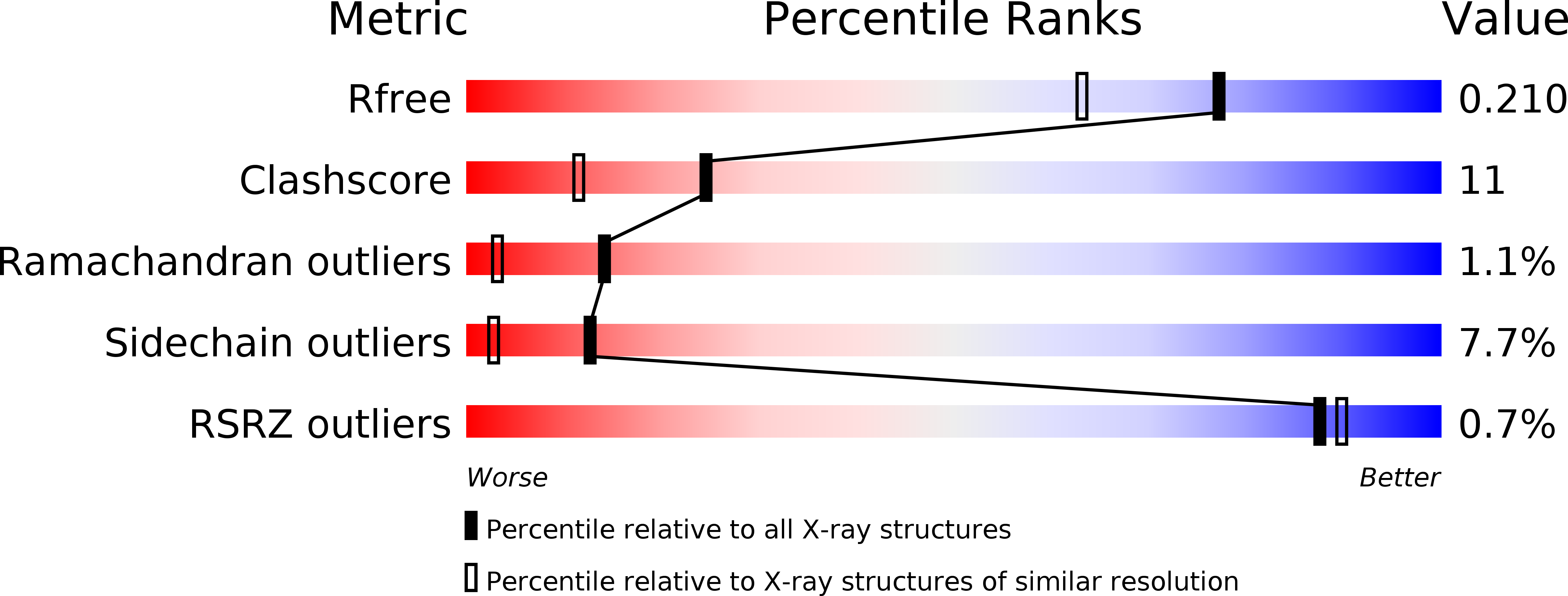
R-Value Free:
0.21
R-Value Work:
0.18
R-Value Observed:
0.18
Space Group:
P 32