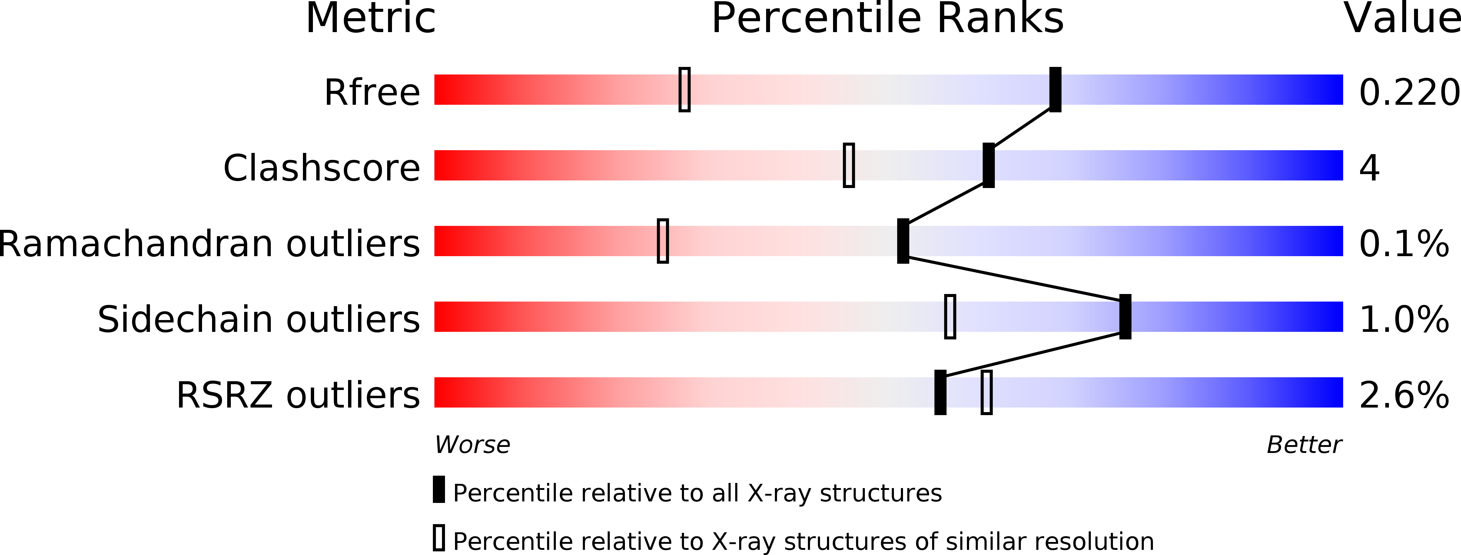
2UVK image
Deposition Date 2007-03-12
Release Date 2007-12-04
Last Version Date 2024-10-23
Entry Detail
PDB ID:
2UVK
Title:
Structure of YjhT
Biological Source:
Source Organism:
Host Organism:
Method Details:
Experimental Method:
Resolution:
1.50 Å
R-Value Free:
0.21
R-Value Work:
0.18
R-Value Observed:
0.18
Space Group:
P 1 21 1
Macromolecular Entities
Structures with similar UniProt ID
Protein Blast
Polymer Type:polypeptide(L)
Molecule:YJHT
Gene (Uniprot):nanM
Chain IDs:A, B
Chain Length:357
Number of Molecules:2
Biological Source:ESCHERICHIA COLI
Modified Residue
Compound ID Chain ID Parent Comp ID Details 2D Image
MSE A MET SELENOMETHIONINE
Primary Citation
Sialic Acid Mutarotation is Catalyzed by the Escherichia Coli Beta-Propeller Protein Yjht.
J.Biol.Chem. 283 4841 ? (2008)
PMID: 18063573 DOI: 10.1074/JBC.M707822200

Abstact

The acquisition of host-derived sialic acid is an important virulence factor for some bacterial pathogens, but in vivo this sugar acid is sequestered in sialoconjugates as the alpha-anomer. In solution, however, sialic acid is present mainly as the beta-anomer, formed by a slow spontaneous mutarotation. We studied the Escherichia coli protein YjhT as a member of a family of uncharacterized proteins present in many sialic acid-utilizing pathogens. This protein is able to accelerate the equilibration of the alpha- and beta-anomers of the sialic acid N-acetylneuraminic acid, thus describing a novel sialic acid mutarotase activity. The structure of this periplasmic protein, solved to 1.5A resolution, reveals a dimeric 6-bladed unclosed beta-propeller, the first of a bacterial Kelch domain protein. Mutagenesis of conserved residues in YjhT demonstrated an important role for Glu-209 and Arg-215 in mutarotase activity. We also present data suggesting that the ability to utilize alpha-N-acetylneuraminic acid released from complex sialoconjugates in vivo provides a physiological advantage to bacteria containing YjhT.

Legend

Protein

Chemical

Disease

Primary Citation of related structures