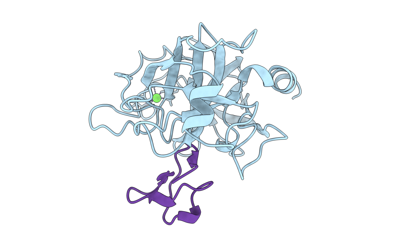
2STA image
Entry Detail
PDB ID:
2STA
Title:
ANIONIC SALMON TRYPSIN IN COMPLEX WITH SQUASH SEED INHIBITOR (CUCURBITA MAXIMA TRYPSIN INHIBITOR I)
Biological Source:
Source Organism:
PDB Version:
Deposition Date:
1998-12-10
Release Date:
2000-01-19
Method Details:
Experimental Method:
Resolution:
1.80 Å
R-Value Free:
0.23
R-Value Work:
0.19
Space Group:
P 21 21 21
Macromolecular Entities
Polymer Type:polypeptide(L)
Description:PROTEIN (TRYPSIN)
Chain IDs:A (auth: E)
Chain Length:222
Number of Molecules:1
Biological Source:Salmo salar
Polymer Type:polypeptide(L)
Description:PROTEIN (TRYPSIN INHIBITOR)
Chain IDs:B (auth: I)
Chain Length:29
Number of Molecules:1
Biological Source:Cucurbita maxima
Ligand Molecules
Primary Citation
High-resolution structures of three new trypsin-squash-inhibitor complexes: a detailed comparison with other trypsins and their complexes.
Acta Crystallogr.,Sect.D 55 139 148 (1999)
PMID: 10089404 DOI: 10.1107/S090744499801052X

Abstact

An anionic trypsin from Atlantic salmon and bovine trypsin have been complexed with the squash-seed inhibitors, CMTI-I (Cucurbita maxima trypsin inhibitor I, P1 Arg) and CPTI-II (Cucurbita pepo trypsin inhibitor II, P1 Lys). The crystal structures of three such complexes have been determined to 1.5-1.8 A resolution and refined to crystallographic R factors ranging from 17.6 to 19.3%. The two anionic salmon-trypsin complexes (ST-CPTI and ST-CMTI) and the bovine-trypsin complex (BT-CPTI) have been compared to other trypsin-inhibitor complexes by means of general structure and primary and secondary binding features. In all three new structures, the primary binding residue of the inhibitor binds to trypsin in the classical manner, but with small differences in the primary and secondary binding patterns. Lysine in CPTI-II binds deeper in the specificity pocket of bovine trypsin than lysine in other known lysine-bovine-trypsin complexes, and anionic salmon trypsin lacks some of the secondary binding interactions found in the complexes formed between squash inhibitors and bovine trypsin. The ST-CMTI complex was formed from the reactive-site-cleaved form of the inhibitor. However, well defined electron density was observed for the P1-P1' peptide bond, together with a hydrogen-bonding pattern virtually identical to those of all serine-protease-protein-inhibitor complexes, indicating a resynthesis of the scissile bond.

Legend

Protein

Chemical

Disease

Primary Citation of related structures