Deposition Date
1998-12-11
Release Date
1998-12-16
Last Version Date
2023-08-30
Entry Detail
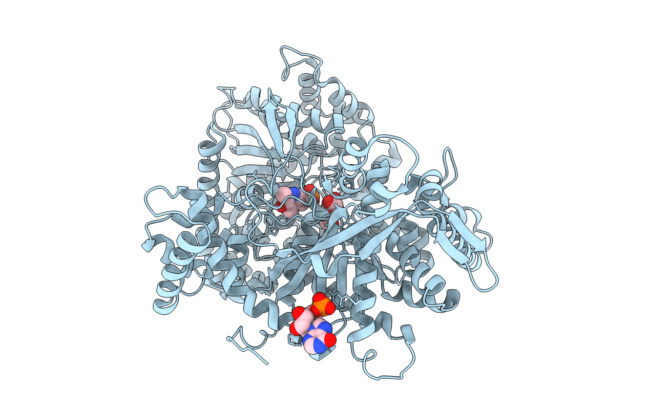
PDB ID:
2SKE
Keywords:
Title:
PYRIDOXAL PHOSPHORYLASE B IN COMPLEX WITH PHOSPHITE, GLUCOSE AND INOSINE-5'-MONOPHOSPHATE
Biological Source:
Source Organism(s):
Oryctolagus cuniculus (Taxon ID: 9986)
Method Details:
Experimental Method:
Resolution:
2.46 Å
R-Value Free:
0.21
R-Value Work:
0.16
R-Value Observed:
0.16
Space Group:
P 43 21 2