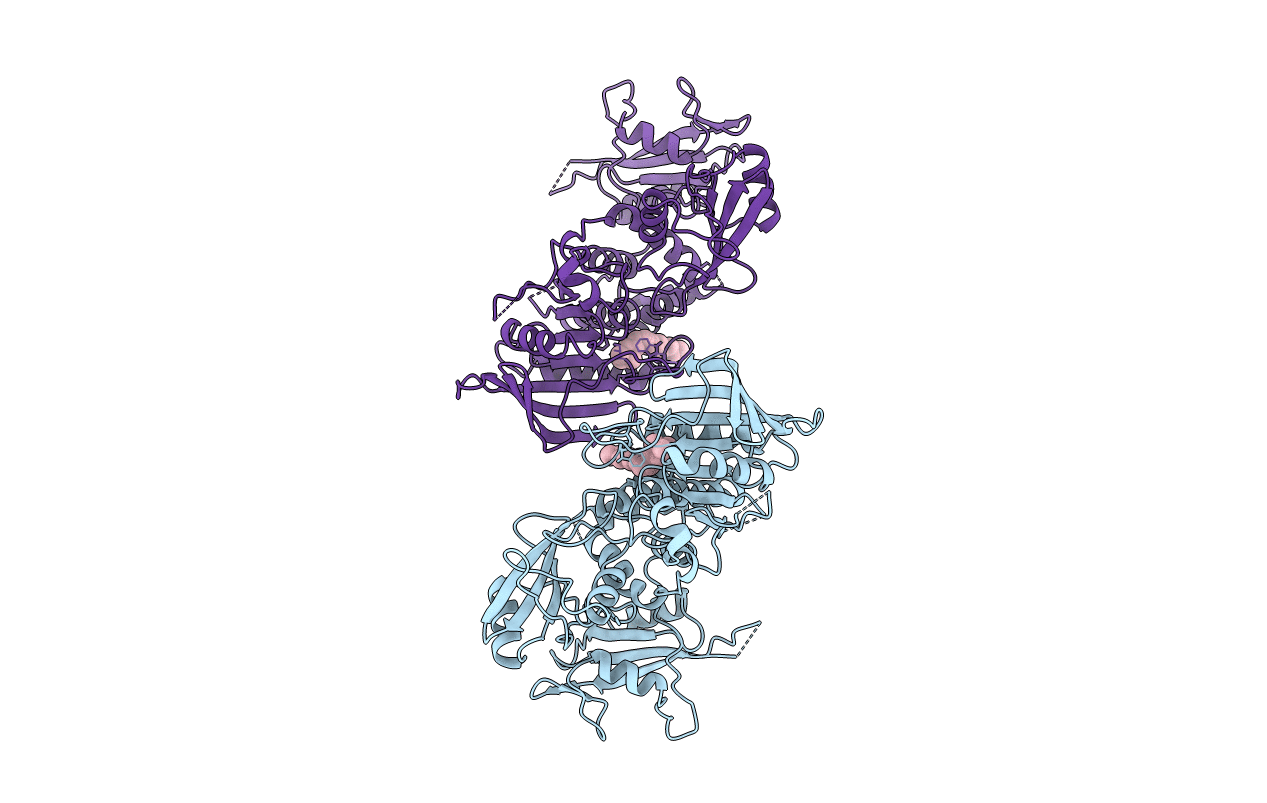
2SHP image
Entry Detail
PDB ID:
2SHP
Title:
TYROSINE PHOSPHATASE SHP-2
Biological Source:
Source Organism:
Host Organism:
PDB Version:
Deposition Date:
1997-12-01
Release Date:
1999-02-16
Method Details:
Experimental Method:
Resolution:
2.00 Å
R-Value Free:
0.27
R-Value Work:
0.19
R-Value Observed:
0.19
Space Group:
P 1 21 1
Macromolecular Entities
Polymer Type:polypeptide(L)
Description:SHP-2
Mutations:T2K, F41L, F513S, DEL(528-593)
Chain IDs:A, B
Chain Length:525
Number of Molecules:2
Biological Source:Homo sapiens
Ligand Molecules
Primary Citation
Crystal structure of the tyrosine phosphatase SHP-2.
Cell(Cambridge,Mass.) 92 441 450 (1998)
PMID: 9491886 DOI: 10.1016/S0092-8674(00)80938-1

Abstact

The structure of the SHP-2 tyrosine phosphatase, determined at 2.0 angstroms resolution, shows how its catalytic activity is regulated by its two SH2 domains. In the absence of a tyrosine-phosphorylated binding partner, the N-terminal SH2 domain binds the phosphatase domain and directly blocks its active site. This interaction alters the structure of the N-SH2 domain, disrupting its phosphopeptide-binding cleft. Conversely, interaction of the N-SH2 domain with phosphopeptide disrupts its phosphatase recognition surface. Thus, the N-SH2 domain is a conformational switch; it either binds and inhibits the phosphatase, or it binds phosphoproteins and activates the enzyme. Recognition of bisphosphorylated ligands by the tandem SH2 domains is an integral element of this switch; the C-terminal SH2 domain contributes binding energy and specificity, but it does not have a direct role in activation.

Legend

Protein

Chemical

Disease

Primary Citation of related structures