2SCU image
Deposition Date 1998-09-24
Release Date 1999-08-02
Last Version Date 2023-12-27
Entry Detail
PDB ID:
2SCU
Keywords:
Title:
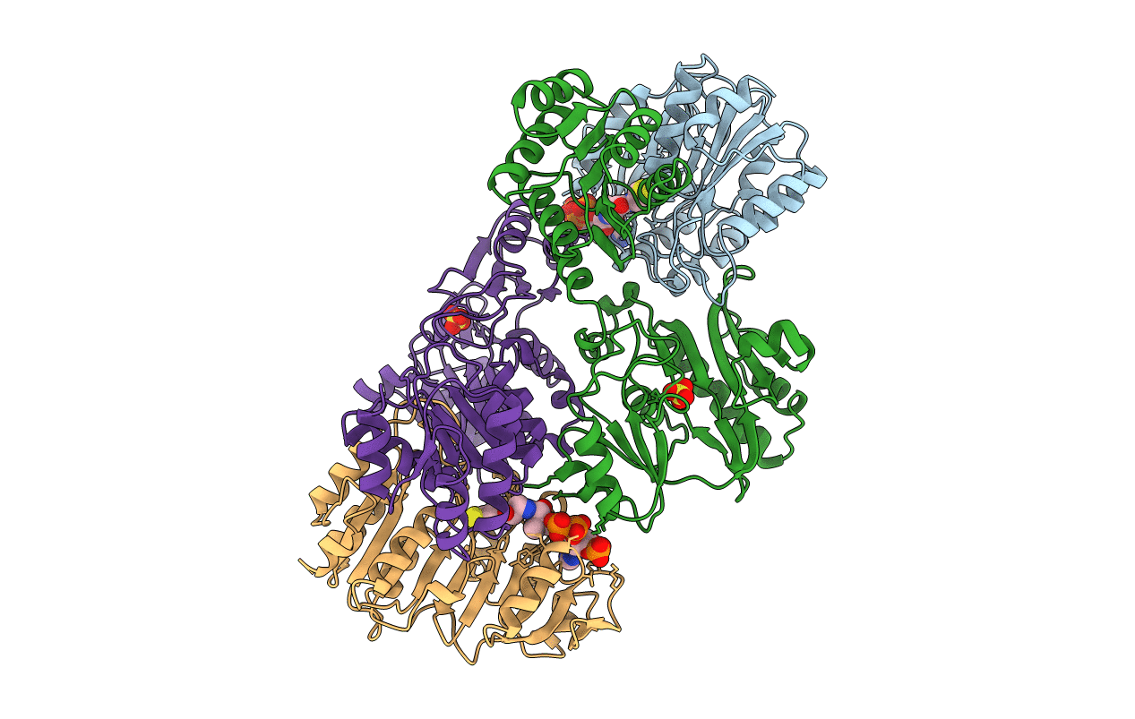
A detailed description of the structure of Succinyl-COA synthetase from Escherichia coli
Biological Source:
Source Organism:
Escherichia coli (Taxon ID: 562)
Host Organism:
Method Details:
Experimental Method:
Resolution:
2.30 Å
R-Value Work:
0.19
Space Group:
P 43 2 2
Macromolecular Entities
Structures with similar UniProt ID
Protein Blast
Polymer Type:polypeptide(L)
Molecule:PROTEIN (SUCCINYL-COA LIGASE)
Chain IDs:A, C (auth: D)
Chain Length:288
Number of Molecules:2
Biological Source:Escherichia coli
Structures with similar UniProt ID
Protein Blast
Polymer Type:polypeptide(L)
Molecule:PROTEIN (SUCCINYL-COA LIGASE)
Chain IDs:B, D (auth: E)
Chain Length:388
Number of Molecules:2
Biological Source:Escherichia coli
Modified Residue
Compound ID Chain ID Parent Comp ID Details 2D Image
NEP A HIS N1-PHOSPHONOHISTIDINE
Primary Citation
A detailed structural description of Escherichia coli succinyl-CoA synthetase.
J.Mol.Biol. 285 1633 1653 (1999)
PMID: 9917402 DOI: 10.1006/jmbi.1998.2324

Abstact

Succinyl-CoA synthetase (SCS) carries out the substrate-level phosphorylation of GDP or ADP in the citric acid cycle. A molecular model of the enzyme from Escherichia coli, crystallized in the presence of CoA, has been refined against data collected to 2.3 A resolution. The crystals are of space group P4322, having unit cell dimensions a=b=98.68 A, c=403.76 A and the data set includes the data measured from 23 crystals. E. coli SCS is an (alphabeta)2-tetramer; there are two copies of each subunit in the asymmetric unit of the crystals. The crystal packing leaves two choices for which pair of alphabeta-dimers form the physiologically relevant tetramer. The copies of the alphabeta-dimer are similar, each having one active site where the phosphorylated histidine residue and the thiol group of CoA are found. CoA is bound in an extended conformation to the nucleotide-binding motif in the N-terminal domain of the alpha-subunit. The phosphoryl group of the phosphorylated histidine residue is positioned at the amino termini of two alpha-helices, one from the C-terminal domain of the alpha-subunit and the other from the C-terminal domain of the beta-subunit. These two domains have similar topologies, despite only 14 % sequence identity. By analogy to other nucleotide-binding proteins, the binding site for the nucleotide may reside in the N-terminal domain of the beta-subunit. If this is so, the catalytic histidine residue would have to move about 35 A to react with the nucleotide.

Legend

Protein

Chemical

Disease

Primary Citation of related structures
Feedback Form
Name
Email
Institute
Feedback