Deposition Date
1991-08-22
Release Date
1993-10-31
Last Version Date
2024-02-21
Entry Detail
PDB ID:
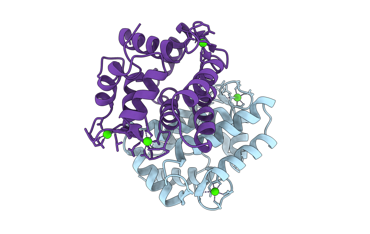
2SCP
Keywords:
Title:
STRUCTURE OF A SARCOPLASMIC CALCIUM-BINDING PROTEIN FROM NEREIS DIVERSICOLOR REFINED AT 2.0 ANGSTROMS RESOLUTION
Biological Source:
Source Organism(s):
Neanthes diversicolor (Taxon ID: 6352)
Method Details:
Experimental Method:
Resolution:
2.00 Å
R-Value Observed:
0.18
Space Group:
P 1 21 1