Deposition Date
2016-03-28
Release Date
2016-05-25
Last Version Date
2024-05-01
Entry Detail
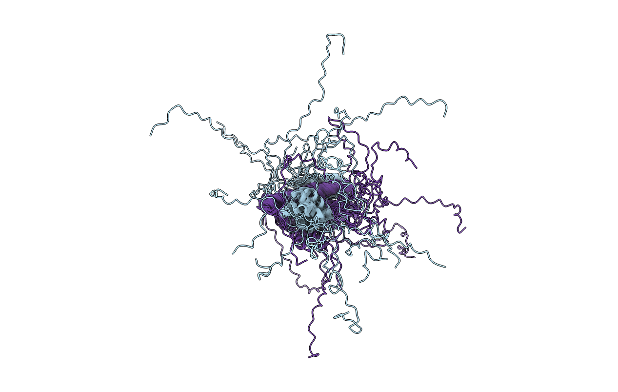
PDB ID:
2RVQ
Keywords:
Title:
Solution structure of the isolated histone H2A-H2B heterodimer
Biological Source:
Source Organism(s):
Homo sapiens (Taxon ID: 9606)
Expression System(s):
Method Details:
Experimental Method:
Conformers Calculated:
10000
Conformers Submitted:
10
Selection Criteria:
structures with the lowest energy