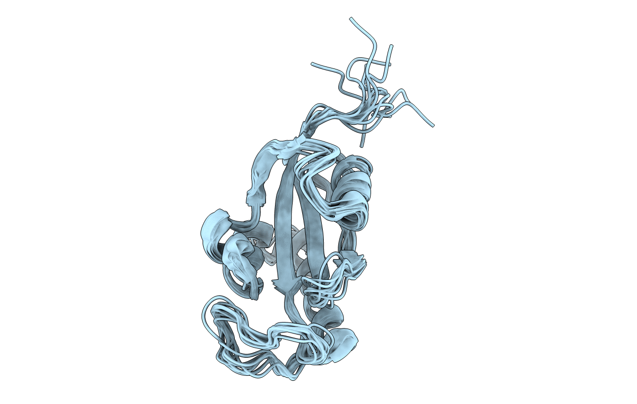
2RUQ image
Deposition Date 2015-01-20
Release Date 2016-01-06
Last Version Date 2024-05-15
Entry Detail
PDB ID:
2RUQ
Keywords:
Title:
solution structure of human Pin1 PPIase mutant C113A
Biological Source:
Source Organism:
Homo sapiens (Taxon ID: 9606)
Host Organism:
Method Details:
Experimental Method:
Conformers Calculated:
50
Conformers Submitted:
10
Selection Criteria:
structures with the lowest energy
Macromolecular Entities
Polymer Type:polypeptide(L)
Molecule:Peptidyl-prolyl cis-trans isomerase NIMA-interacting 1
Gene (Uniprot):PIN1
Mutations:C113A
Chain IDs:A
Chain Length:117
Number of Molecules:1
Biological Source:Homo sapiens
Ligand Molecules
Primary Citation
Allosteric Breakage of the Hydrogen Bond within the Dual-Histidine Motif in the Active Site of Human Pin1 PPIase
Biochemistry 54 5242 5253 (2015)
PMID: 26226559 DOI: 10.1021/acs.biochem.5b00606

Abstact

Intimate cooperativity among active site residues in enzymes is a key factor for regulating elaborate reactions that would otherwise not occur readily. Peptidyl-prolyl cis-trans isomerase NIMA-interacting 1 (Pin1) is the phosphorylation-dependent cis-trans peptidyl-prolyl isomerase (PPIase) that specifically targets phosphorylated Ser/Thr-Pro motifs. Residues C113, H59, H157, and T152 form a hydrogen bond network in the active site, as in the noted connection. Theoretical studies have shown that protonation to thiolate C113 leads to rearrangement of this hydrogen bond network, with switching of the tautomeric states of adjacent histidines (H59 and H157) [Barman, A., and Hamelberg, D. (2014) Biochemistry 53, 3839-3850]. This is called the "dual-histidine motif". Here, C113A and C113S Pin1 mutants were found to alter the protonation states of H59 according to the respective residue type replaced at C113, and the mutations resulted in disruption of the hydrogen bond within the dual-histidine motif. In the C113A mutant, H59 was observed to be in exchange between ε- and δ-tautomers, which widened the entrance of the active site cavity, as seen by an increase in the distance between residues A113 and S154. The C113S mutant caused H59 to exchange between the ε-tautomer and imidazolium while not changing the active site structure. Moreover, the imidazole ring orientations of H59 and H157 were changed in the C113S mutant. These results demonstrated that a mutation at C113 modulates the hydrogen bond network dynamics. Thus, C113 acts as a pivot to drive the concerted function among the residues in the hydrogen bond network, as theoretically predicted.

Legend

Protein

Chemical

Disease

Primary Citation of related structures