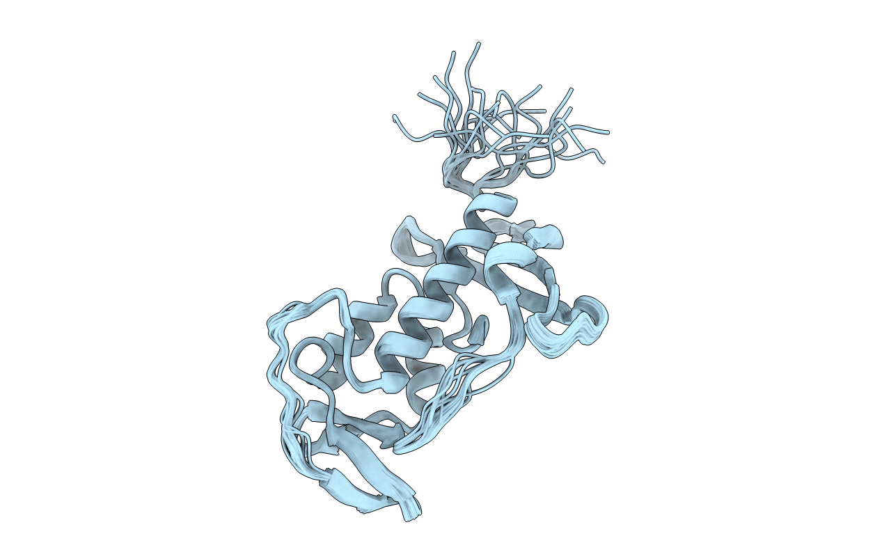
Abstact
In Bacillus subtilis, LytE, LytF, CwlS, and CwlO are vegetative autolysins, DL-endopeptidases in the NlpC/P60 family, and play essential roles in cell growth and separation. IseA (YoeB) is a proteinaceous inhibitor against the DL-endopeptidases, peptidoglycan hydrolases. Overexpression of IseA caused significantly long chained cell morphology, because IseA inhibits the cell separation DL-endopeptidases post-translationally. Here, we report the first three-dimensional structure of IseA, determined by NMR spectroscopy. The structure includes a single domain consisting of three α-helices, one 3(10)-helix, and eight β-strands, which is a novel fold like a "hacksaw." Noteworthy is a dynamic loop between β4 and the 3(10)-helix, which resembles a "blade." The electrostatic potential distribution shows that most of the surface is positively charged, but the region around the loop is negatively charged. In contrast, the LytF active-site cleft is expected to be positively charged. NMR chemical shift perturbation of IseA interacting with LytF indicated that potential interaction sites are located around the loop. Furthermore, the IseA mutants D100K/D102K and G99P/G101P at the loop showed dramatic loss of inhibition activity against LytF, compared with wild-type IseA, indicating that the β4-3(10) loop plays an important role in inhibition. Moreover, we built a complex structure model of IseA-LytF by docking simulation, suggesting that the β4-3(10) loop of IseA gets stuck deep in the cleft of LytF, and the active site is occluded. These results suggest a novel inhibition mechanism of the hacksaw-like structure, which is different from known inhibitor proteins, through interactions around the characteristic loop regions with the active-site cleft of enzymes.