2RR7 image
Deposition Date 2010-06-04
Release Date 2011-06-08
Last Version Date 2024-05-01
Entry Detail
PDB ID:
2RR7
Keywords:
Title:
Microtubule Binding Domain of DYNEIN-C
Biological Source:
Source Organism:
Host Organism:
Method Details:
Experimental Method:
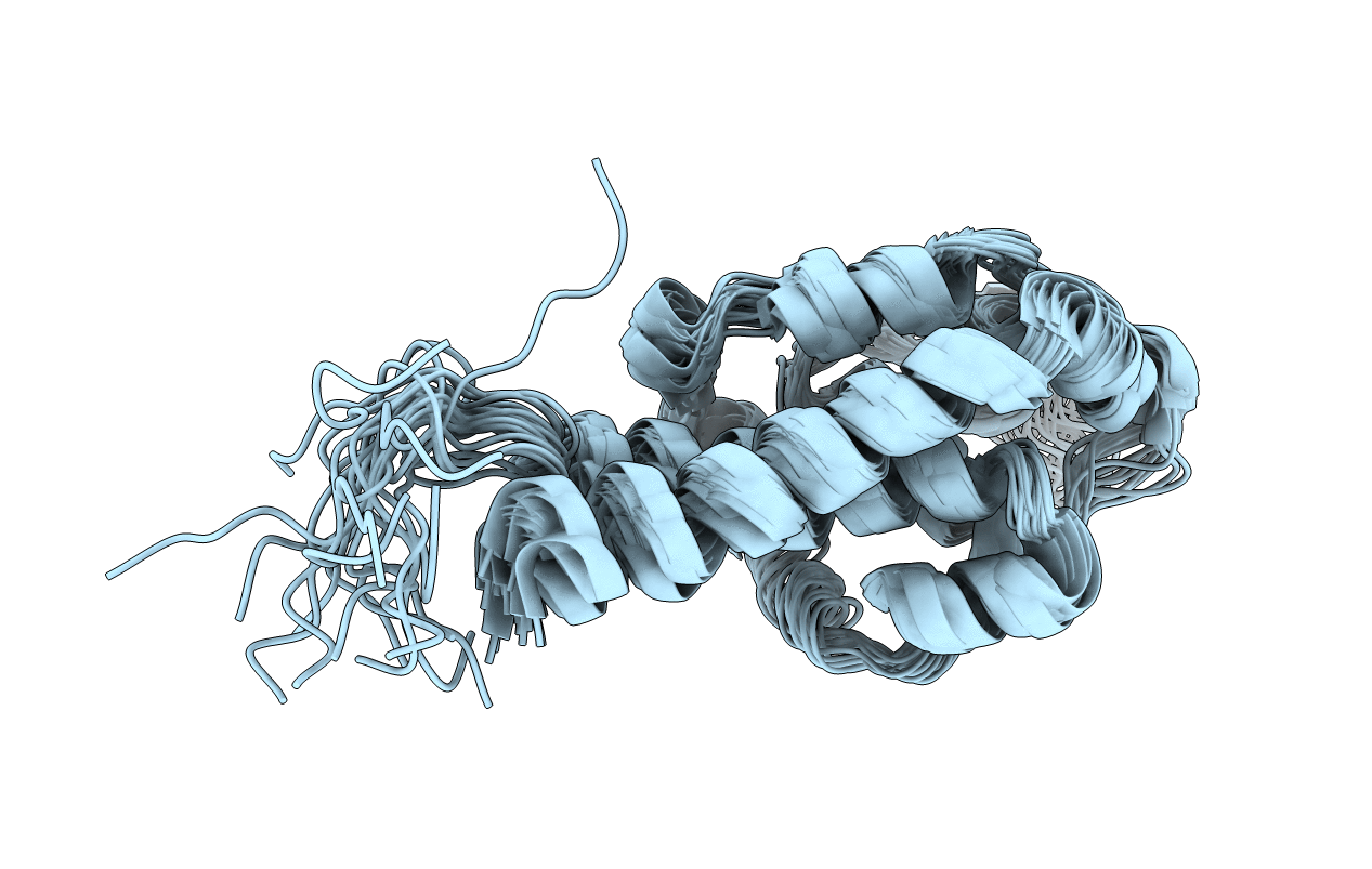
Conformers Calculated:
200
Conformers Submitted:
20
Selection Criteria:
target function
Macromolecular Entities
Polymer Type:polypeptide(L)
Molecule:Dynein heavy chain 9
Gene (Uniprot):DHC9
Chain IDs:A
Chain Length:155
Number of Molecules:1
Biological Source:Chlamydomonas reinhardtii
Ligand Molecules
Primary Citation
Structure of the microtubule-binding domain of flagellar dynein
Structure 22 1628 1638 (2014)
PMID: 25450768 DOI: 10.1016/j.str.2014.08.021

Abstact

Flagellar dyneins are essential microtubule motors in eukaryotes, as they drive the beating motions of cilia and flagella. Unlike myosin and kinesin motors, the track binding mechanism of dyneins and the regulation between the strong and weak binding states remain obscure. Here we report the solution structure of the microtubule-binding domain of flagellar dynein-c/DHC9 (dynein-c MTBD). The structure reveals a similar overall helix-rich fold to that of the MTBD of cytoplasmic dynein (cytoplasmic MTBD), but dynein-c MTBD has an additional flap, consisting of an antiparallel b sheet. The flap is positively charged and highly flexible. Despite the structural similarity to cytoplasmic MTBD, dynein-c MTBD shows only a small change in the microtubule- binding affinity depending on the registry change of coiled coil-sliding, whereby lacks the apparent strong binding state. The surface charge distribution of dynein-c MTBD also differs from that of cytoplasmic MTBD, which suggests a difference in the microtubule-binding mechanism.

Legend

Protein

Chemical

Disease

Primary Citation of related structures