Deposition Date
2009-11-17
Release Date
2010-11-24
Last Version Date
2024-05-01
Entry Detail
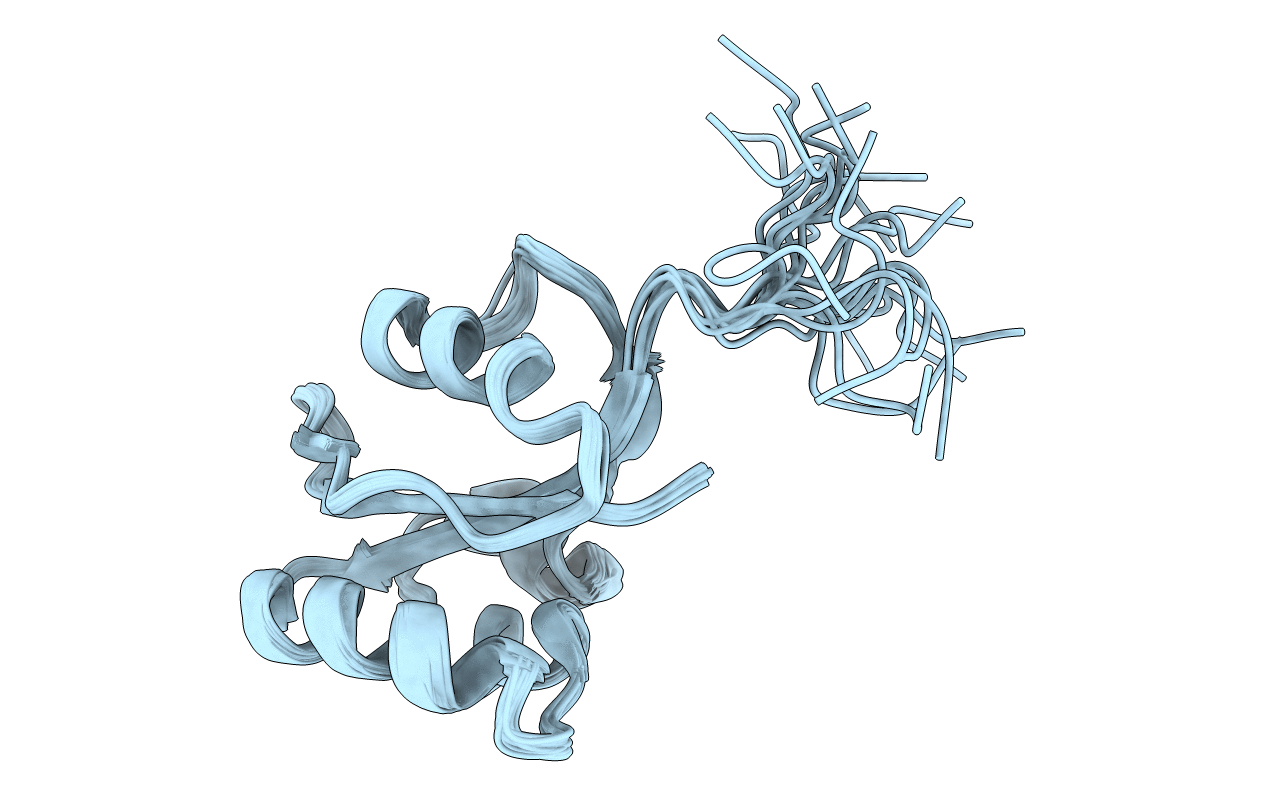
PDB ID:
2RQS
Keywords:
Title:
3D structure of Pin from the psychrophilic archeon Cenarcheaum symbiosum (CsPin)
Biological Source:
Source Organism(s):
Cenarchaeum symbiosum (Taxon ID: 46770)
Expression System(s):
Method Details:
Experimental Method:
Conformers Calculated:
200
Conformers Submitted:
20
Selection Criteria:
structures with the lowest energy