2RPW image
Deposition Date 2008-11-08
Release Date 2009-03-24
Last Version Date 2024-05-29
Entry Detail
PDB ID:
2RPW
Title:
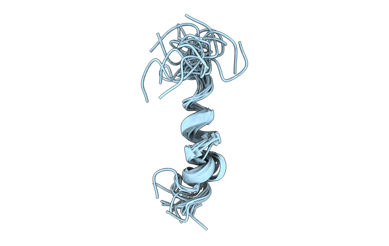
Structure of a peptide derived from H+-V-ATPase subunit a
Biological Source:
Source Organism:
(Taxon ID: )
Method Details:
Experimental Method:
Conformers Calculated:
100
Conformers Submitted:
20
Selection Criteria:
structures with the lowest energy
Macromolecular Entities
Polymer Type:polypeptide(L)
Molecule:25 meric peptide from V-type proton ATPase subunit a, vacuolar isoform
Gene (Uniprot):VPH1
Chain IDs:A (auth: X)
Chain Length:25
Number of Molecules:1
Biological Source:
Ligand Molecules
Primary Citation
Structural properties of a peptide derived from H(+)-V-ATPase subunit a
Biochim.Biophys.Acta 1788 1204 1212 (2009)
PMID: 19249284 DOI: 10.1016/j.bbamem.2009.02.015

Abstact

The 3D structure of a peptide derived from the putative transmembrane segment 7 (TM7) of subunit a from H(+)-V-ATPase from Saccharomyces cerevisiae has been determined by solution state NMR in SDS. A stable helix is formed from L736 up to and including Q745, the lumenal half of the putative TM7. The helical region extends well beyond A738, as was previously suggested based on NMR studies of a similar peptide in DMSO. The pKa of both histidine residues that are important for proton transport was measured in water and in SDS. The differences that are found demonstrate that the histidine residues interact with the SDS polar heads. In detergent, circular dichroism data indicate that the secondary structure of the peptide depends on the pH and the type of detergent used. Using solid-state NMR, it is shown that the peptide is immobile in phospholipid bilayers, which means that it is probably not a single transmembrane helix in these samples. The environment is important for the structure of TM7, so in subunit a it is probably held in place by the other transmembrane helices of this subunit.

Legend

Protein

Chemical

Disease

Primary Citation of related structures