Deposition Date
1988-07-06
Release Date
1989-10-15
Last Version Date
2024-11-20
Entry Detail
PDB ID:
2RNT
Keywords:
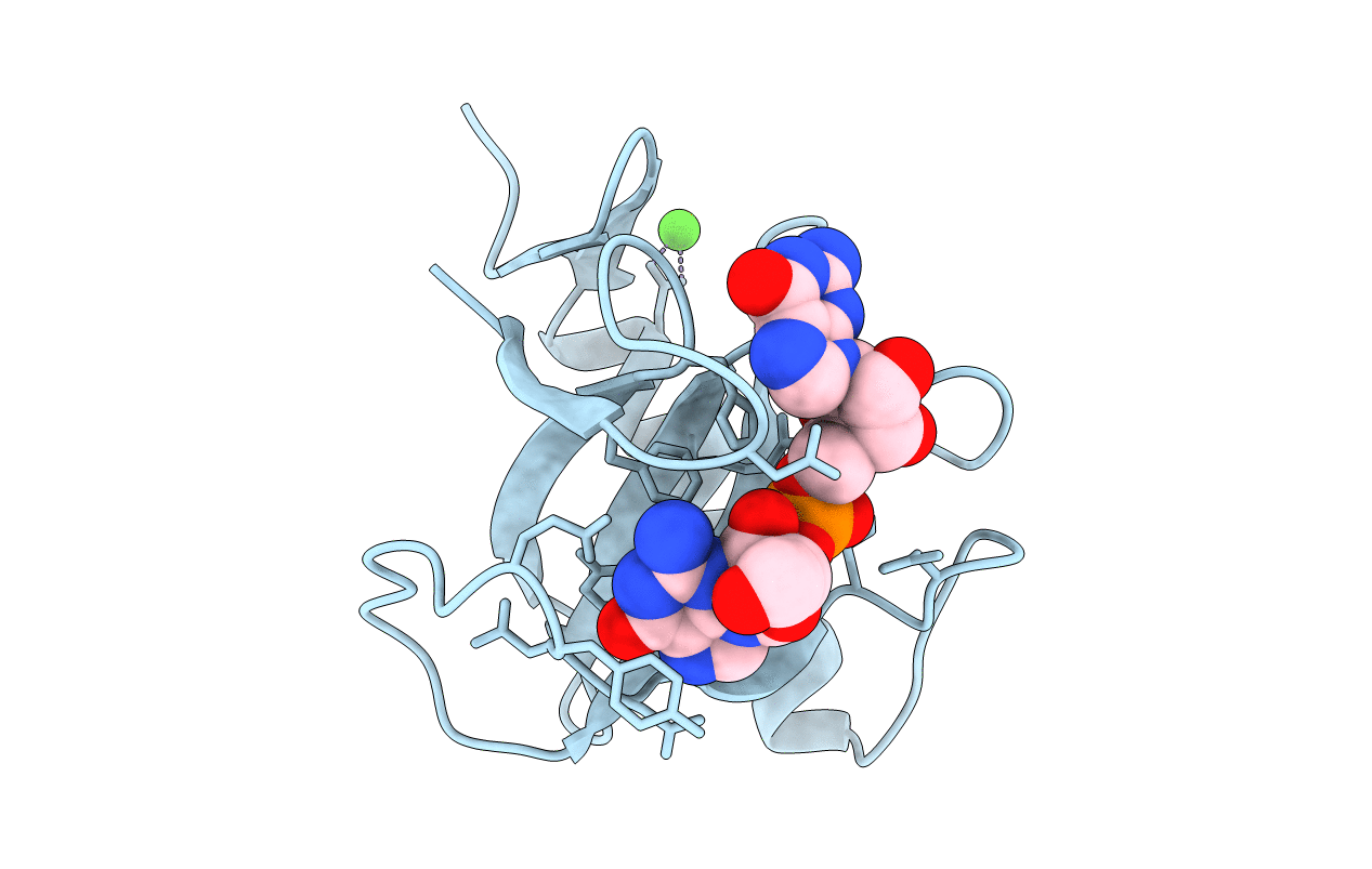
Title:
THREE-DIMENSIONAL STRUCTURE OF RIBONUCLEASE T1 COMPLEXED WITH GUANYLYL-2(PRIME),5(PRIME)-GUANOSINE AT 1.8 ANGSTROMS RESOLUTION
Biological Source:
Source Organism(s):
Aspergillus oryzae (Taxon ID: 5062)
Method Details:
Experimental Method:
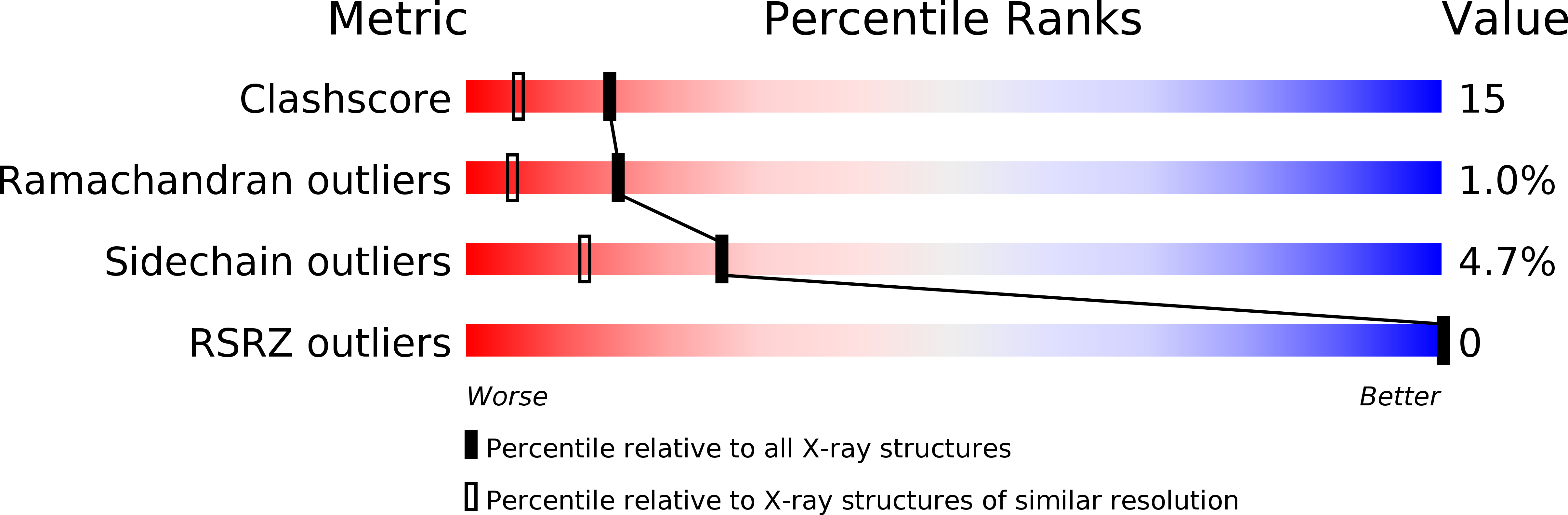
Resolution:
1.80 Å
R-Value Observed:
0.14
Space Group:
P 21 21 21